 Movie
Movie Controller

[English] 日本語
 Yorodumi
- PDB-3mjg: The structure of a platelet derived growth factor receptor complex -
+ Open data

- Basic information
| Entry | Database: PDB / ID: 3mjg | |||||||||
|---|---|---|---|---|---|---|---|---|---|---|
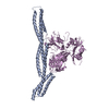
| Title | The structure of a platelet derived growth factor receptor complex | |||||||||
 Components Components |
| |||||||||
 Keywords Keywords | Hormone/Transferase / protein-protein complex / growth factor-receptor complex / Transferase-Hormone complex / Hormone-Transferase complex | |||||||||
| Function / homology |  Function and homology information Function and homology informationmetanephric glomerular mesangial cell development / positive regulation of vascular associated smooth muscle cell dedifferentiation /  platelet-derived growth factor complex / positive regulation of metanephric mesenchymal cell migration / negative regulation of phosphatidylinositol biosynthetic process / platelet activating factor receptor activity / platelet-derived growth factor receptor activity / platelet-derived growth factor beta-receptor activity / cell migration involved in coronary angiogenesis / metanephric glomerular mesangial cell proliferation involved in metanephros development ...metanephric glomerular mesangial cell development / positive regulation of vascular associated smooth muscle cell dedifferentiation / platelet-derived growth factor complex / positive regulation of metanephric mesenchymal cell migration / negative regulation of phosphatidylinositol biosynthetic process / platelet activating factor receptor activity / platelet-derived growth factor receptor activity / platelet-derived growth factor beta-receptor activity / cell migration involved in coronary angiogenesis / metanephric glomerular mesangial cell proliferation involved in metanephros development / positive regulation of glomerular filtration / positive regulation of metanephric mesenchymal cell migration by platelet-derived growth factor receptor-beta signaling pathway / smooth muscle cell chemotaxis / metanephric glomerular capillary formation / cellular response to mycophenolic acid / superoxide-generating NADPH oxidase activator activity / negative regulation of vascular associated smooth muscle cell differentiation / cell migration involved in vasculogenesis / protein kinase C signaling / aorta morphogenesis / positive regulation of hyaluronan biosynthetic process / positive regulation of cell proliferation by VEGF-activated platelet derived growth factor receptor signaling pathway / platelet-derived growth factor binding / retina vasculature development in camera-type eye / vascular endothelial growth factor binding / cardiac myofibril assembly / phosphatidylinositol metabolic process / positive regulation of glomerular mesangial cell proliferation / positive regulation of chemotaxis / interleukin-18-mediated signaling pathway / Signaling by PDGF / paracrine signaling / platelet-derived growth factor receptor binding / positive regulation of vascular associated smooth muscle cell migration / positive regulation of cell-substrate adhesion / positive regulation of DNA biosynthetic process / cellular response to platelet-derived growth factor stimulus / positive regulation of smooth muscle cell migration / positive regulation of calcium ion import / chemoattractant activity / platelet-derived growth factor receptor-beta signaling pathway / monocyte chemotaxis / positive regulation of phosphoprotein phosphatase activity / positive regulation of cell division / platelet-derived growth factor receptor signaling pathway / Non-integrin membrane-ECM interactions / negative regulation of platelet activation / embryonic placenta development / positive regulation of blood vessel endothelial cell migration / positive regulation of phospholipase C activity / positive regulation of calcium-mediated signaling / positive regulation of protein autophosphorylation / positive regulation of vascular associated smooth muscle cell proliferation / collagen binding / reactive oxygen species metabolic process / positive regulation of endothelial cell proliferation / cell chemotaxis / Downstream signal transduction / lysosomal lumen / positive regulation of mitotic nuclear division / negative regulation of miRNA transcription / platelet alpha granule lumen / negative regulation of protein binding / regulation of actin cytoskeleton organization / positive regulation of smooth muscle cell proliferation / growth factor activity / positive regulation of MAP kinase activity / receptor protein-tyrosine kinase / cellular response to growth factor stimulus / response to wounding / positive regulation of miRNA transcription / Golgi lumen / peptidyl-tyrosine phosphorylation / cell surface receptor protein tyrosine kinase signaling pathway / Constitutive Signaling by Aberrant PI3K in Cancer / positive regulation of reactive oxygen species metabolic process / positive regulation of fibroblast proliferation / positive regulation of peptidyl-tyrosine phosphorylation / Platelet degranulation / PIP3 activates AKT signaling / gene expression / heart development / PI5P, PP2A and IER3 Regulate PI3K/AKT Signaling / cytoplasmic vesicle / RAF/MAP kinase cascade / basolateral plasma membrane / protein tyrosine kinase activity / collagen-containing extracellular matrix / positive regulation of MAPK cascade / protein autophosphorylation / positive regulation of phosphatidylinositol 3-kinase/protein kinase B signal transduction / positive regulation of ERK1 and ERK2 cascade / receptor complex / protein kinase activity / positive regulation of cell migration / apical plasma membrane / protein heterodimerization activity / endoplasmic reticulum lumen / Golgi membrane / protein phosphorylation platelet-derived growth factor complex / positive regulation of metanephric mesenchymal cell migration / negative regulation of phosphatidylinositol biosynthetic process / platelet activating factor receptor activity / platelet-derived growth factor receptor activity / platelet-derived growth factor beta-receptor activity / cell migration involved in coronary angiogenesis / metanephric glomerular mesangial cell proliferation involved in metanephros development ...metanephric glomerular mesangial cell development / positive regulation of vascular associated smooth muscle cell dedifferentiation / platelet-derived growth factor complex / positive regulation of metanephric mesenchymal cell migration / negative regulation of phosphatidylinositol biosynthetic process / platelet activating factor receptor activity / platelet-derived growth factor receptor activity / platelet-derived growth factor beta-receptor activity / cell migration involved in coronary angiogenesis / metanephric glomerular mesangial cell proliferation involved in metanephros development / positive regulation of glomerular filtration / positive regulation of metanephric mesenchymal cell migration by platelet-derived growth factor receptor-beta signaling pathway / smooth muscle cell chemotaxis / metanephric glomerular capillary formation / cellular response to mycophenolic acid / superoxide-generating NADPH oxidase activator activity / negative regulation of vascular associated smooth muscle cell differentiation / cell migration involved in vasculogenesis / protein kinase C signaling / aorta morphogenesis / positive regulation of hyaluronan biosynthetic process / positive regulation of cell proliferation by VEGF-activated platelet derived growth factor receptor signaling pathway / platelet-derived growth factor binding / retina vasculature development in camera-type eye / vascular endothelial growth factor binding / cardiac myofibril assembly / phosphatidylinositol metabolic process / positive regulation of glomerular mesangial cell proliferation / positive regulation of chemotaxis / interleukin-18-mediated signaling pathway / Signaling by PDGF / paracrine signaling / platelet-derived growth factor receptor binding / positive regulation of vascular associated smooth muscle cell migration / positive regulation of cell-substrate adhesion / positive regulation of DNA biosynthetic process / cellular response to platelet-derived growth factor stimulus / positive regulation of smooth muscle cell migration / positive regulation of calcium ion import / chemoattractant activity / platelet-derived growth factor receptor-beta signaling pathway / monocyte chemotaxis / positive regulation of phosphoprotein phosphatase activity / positive regulation of cell division / platelet-derived growth factor receptor signaling pathway / Non-integrin membrane-ECM interactions / negative regulation of platelet activation / embryonic placenta development / positive regulation of blood vessel endothelial cell migration / positive regulation of phospholipase C activity / positive regulation of calcium-mediated signaling / positive regulation of protein autophosphorylation / positive regulation of vascular associated smooth muscle cell proliferation / collagen binding / reactive oxygen species metabolic process / positive regulation of endothelial cell proliferation / cell chemotaxis / Downstream signal transduction / lysosomal lumen / positive regulation of mitotic nuclear division / negative regulation of miRNA transcription / platelet alpha granule lumen / negative regulation of protein binding / regulation of actin cytoskeleton organization / positive regulation of smooth muscle cell proliferation / growth factor activity / positive regulation of MAP kinase activity / receptor protein-tyrosine kinase / cellular response to growth factor stimulus / response to wounding / positive regulation of miRNA transcription / Golgi lumen / peptidyl-tyrosine phosphorylation / cell surface receptor protein tyrosine kinase signaling pathway / Constitutive Signaling by Aberrant PI3K in Cancer / positive regulation of reactive oxygen species metabolic process / positive regulation of fibroblast proliferation / positive regulation of peptidyl-tyrosine phosphorylation / Platelet degranulation / PIP3 activates AKT signaling / gene expression / heart development / PI5P, PP2A and IER3 Regulate PI3K/AKT Signaling / cytoplasmic vesicle / RAF/MAP kinase cascade / basolateral plasma membrane / protein tyrosine kinase activity / collagen-containing extracellular matrix / positive regulation of MAPK cascade / protein autophosphorylation / positive regulation of phosphatidylinositol 3-kinase/protein kinase B signal transduction / positive regulation of ERK1 and ERK2 cascade / receptor complex / protein kinase activity / positive regulation of cell migration / apical plasma membrane / protein heterodimerization activity / endoplasmic reticulum lumen / Golgi membrane / protein phosphorylationSimilarity search - Function | |||||||||
| Biological species |  Homo sapiens (human) Homo sapiens (human) | |||||||||
| Method | X-RAY DIFFRACTION / SYNCHROTRON / MOLECULAR REPLACEMENT / Resolution: 2.3 Å | |||||||||
 Authors Authors | Shim, A.H.R. / He, X. | |||||||||
 Citation Citation | Journal: Proc.Natl.Acad.Sci.USA / Year: 2010 Title: Structures of a platelet-derived growth factor/propeptide complex and a platelet-derived growth factor/receptor complex. Authors: Shim, A.H. / Liu, H. / Focia, P.J. / Chen, X. / Lin, P.C. / He, X. | |||||||||
| History |
|
- Structure visualization
| Structure viewer | Molecule: MolmilJmol/JSmol |
|---|
- Downloads & links
-Download
| PDBx/mmCIF format | 3mjg.cif.gz | 198.9 KB | Display | PDBx/mmCIF format |
|---|---|---|---|---|
| PDB format | pdb3mjg.ent.gz | 154.5 KB | Display | PDB format |
| PDBx/mmJSON format | 3mjg.json.gz | Tree view | PDBx/mmJSON format | |
| Others |  Other downloads Other downloads |
-Validation report
| Arichive directory | https://data.pdbj.org/pub/pdb/validation_reports/mj/3mjgftp://data.pdbj.org/pub/pdb/validation_reports/mj/3mjg | HTTPS FTP |
|---|
-Related structure data
| Related structure data |  3mjkC 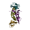 1fltS 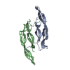 1pdgS S: Starting model for refinement C: citing same article ( |
|---|---|
| Similar structure data |


-Links
 PDBj
PDBj


- Assembly
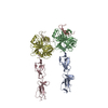
| Deposited unit | 
| ||||||||
|---|---|---|---|---|---|---|---|---|---|
| 1 |
| ||||||||
| 2 | 
| ||||||||
| Unit cell |
|
-Components
| #1: Protein | / PDGF subunit B / Platelet-derived growth factor B chain / Platelet-derived growth factor beta ...PDGF subunit B / Platelet-derived growth factor B chain / Platelet-derived growth factor beta polypeptide / PDGF-2 / Proto-oncogene c-Sis Mass: 19596.301 Da / Num. of mol.: 2 / Fragment: UNP residues 21-185 Source method: isolated from a genetically manipulated source Source: (gene. exp.) Homo sapiens (human) / Gene: PDGF2, PDGFB, SIS / Cell line (production host): Embryonic Kidney-293 cells / Production host: Homo sapiens / References: UniProt: P01127#2: Protein | Mass: 32493.354 Da / Num. of mol.: 2 / Fragment: UNP residues 33-314 Source method: isolated from a genetically manipulated source Source: (gene. exp.) Homo sapiens (human) / Gene: PDGFRB / Cell line (production host): Embryonic Kidney-293 cells / Production host: Homo sapiensReferences: UniProt: P09619, receptor protein-tyrosine kinase#3: Sugar | ChemComp-NDG / | N-Acetylglucosamine  Type: D-saccharide, alpha linking / Mass: 221.208 Da / Num. of mol.: 1 Type: D-saccharide, alpha linking / Mass: 221.208 Da / Num. of mol.: 1Source method: isolated from a genetically manipulated source Formula: C8H15NO6 #4: Sugar | ChemComp-NAG / N-Acetylglucosamine  Type: D-saccharide, beta linking / Mass: 221.208 Da / Num. of mol.: 13 Type: D-saccharide, beta linking / Mass: 221.208 Da / Num. of mol.: 13Source method: isolated from a genetically manipulated source Formula: C8H15NO6 #5: Water | ChemComp-HOH / | Water Mass: 18.015 Da / Num. of mol.: 1170 / Source method: isolated from a natural source / Formula: H2O Mass: 18.015 Da / Num. of mol.: 1170 / Source method: isolated from a natural source / Formula: H2O |
|---|
-Experimental details
-Experiment
| Experiment | Method: X-RAY DIFFRACTION / Number of used crystals: 1 |
|---|
- Sample preparation
| Crystal | Density Matthews: 2.9 Å3/Da / Density % sol: 57.62 % |
|---|---|
| Crystal grow | Temperature: 295 K / Method: vapor diffusion, sitting drop / pH: 8 Details: 0.84 M (NH4)2HPO4, 0.1 M imidazole, pH 8.0, VAPOR DIFFUSION, SITTING DROP, temperature 295K |
-Data collection
| Diffraction | Mean temperature: 100 K |
|---|---|
| Diffraction source | Source: SYNCHROTRON / Site: APS  / Beamline: 21-ID-D / Wavelength: 0.9786 Å / Beamline: 21-ID-D / Wavelength: 0.9786 Å |
| Detector | Type: MAR scanner 300 mm plate / Detector: IMAGE PLATE / Date: Jun 15, 2008 |
| Radiation | Monochromator: SAGITALLY FOCUSED Si(111) / Protocol: SINGLE WAVELENGTH / Monochromatic (M) / Laue (L): M / Scattering type: x-ray |
| Radiation wavelength | Wavelength: 0.9786 Å / Relative weight: 1 |
| Reflection | Resolution: 2.3→44.24 Å / Num. all: 55432 / Num. obs: 54767 / % possible obs: 98.8 % / Observed criterion σ(F): 0 / Observed criterion σ(I): 0 / Redundancy: 4 % / Biso Wilson estimate: 36 Å2 / Rmerge(I) obs: 0.078 / Rsym value: 0.062 / Net I/σ(I): 14.3 |
| Reflection shell | Resolution: 2.3→2.4 Å / Redundancy: 4 % / Rmerge(I) obs: 0.327 / Mean I/σ(I) obs: 3.3 / Num. unique all: 8466 / Rsym value: 0.426 / % possible all: 97.7 |
- Processing
| Software |
| |||||||||||||||||||||||||
|---|---|---|---|---|---|---|---|---|---|---|---|---|---|---|---|---|---|---|---|---|---|---|---|---|---|---|
| Refinement | Method to determine structure: MOLECULAR REPLACEMENT Starting model: PDB entries 1pdg, 1flt Resolution: 2.3→44.24 Å / Rfactor Rfree error: 0.005 / Data cutoff high absF: 2835260.48 / Data cutoff low absF: 0 / Isotropic thermal model: RESTRAINED / Cross valid method: THROUGHOUT / σ(F): 0 / Stereochemistry target values: Engh & Huber / Details: BULK SOLVENT MODEL USED
| |||||||||||||||||||||||||
| Solvent computation | Solvent model: FLAT MODEL / Bsol: 61.54 Å2 / ksol: 0.272531 e/Å3 | |||||||||||||||||||||||||
| Displacement parameters | Biso mean: 67.4 Å2
| |||||||||||||||||||||||||
| Refine analyze |
| |||||||||||||||||||||||||
| Refinement step | Cycle: LAST / Resolution: 2.3→44.24 Å
| |||||||||||||||||||||||||
| Refine LS restraints |
| |||||||||||||||||||||||||
| Refine LS restraints NCS | NCS model details: NONE | |||||||||||||||||||||||||
| LS refinement shell | Resolution: 2.3→2.44 Å / Rfactor Rfree error: 0.017 / Total num. of bins used: 6
| |||||||||||||||||||||||||
| Xplor file |
|
