 Movie
Movie Controller


+ Open data

- Basic information
| Entry | Database: PDB / ID: 3mi9 | ||||||
|---|---|---|---|---|---|---|---|
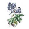
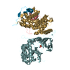
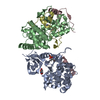
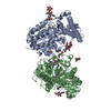
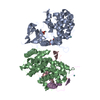
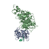
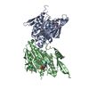
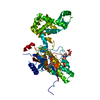
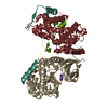
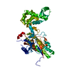
| Title | Crystal structure of HIV-1 Tat complexed with human P-TEFb | ||||||
 Components Components |
| ||||||
 Keywords Keywords |  PROTEIN BINDING / P-TEFb / Tat / HIV-1 PROTEIN BINDING / P-TEFb / Tat / HIV-1 | ||||||
| Function / homology |  Function and homology informationtrans-activation response element binding / P-TEFb complex / Interactions of Tat with host cellular proteins / 7SK snRNA binding / protein serine/threonine phosphatase inhibitor activity / positive regulation of viral transcription / cyclin/CDK positive transcription elongation factor complex / regulation of muscle cell differentiation / regulation of mRNA 3'-end processing / positive regulation of protein localization to chromatin ...trans-activation response element binding / P-TEFb complex / Interactions of Tat with host cellular proteins / 7SK snRNA binding / protein serine/threonine phosphatase inhibitor activity / positive regulation of viral transcription / cyclin/CDK positive transcription elongation factor complex / regulation of muscle cell differentiation / regulation of mRNA 3'-end processing / positive regulation of protein localization to chromatin / modulation by virus of host chromatin organization / symbiont-mediated suppression of host translation initiation / nucleus localization / evasion of host immune response / molecular sequestering activity / host cell nucleolus / actinin binding / cyclin-dependent protein serine/threonine kinase activator activity / positive regulation by host of viral transcription / RNA polymerase binding / positive regulation of DNA-templated transcription, elongation / negative regulation of protein localization to chromatin / [RNA-polymerase]-subunit kinase / transcription elongation-coupled chromatin remodeling / replication fork processing / negative regulation of peptidyl-threonine phosphorylation / regulation of cyclin-dependent protein serine/threonine kinase activity / cellular response to cytokine stimulus / Pausing and recovery of Tat-mediated HIV elongation / Tat-mediated HIV elongation arrest and recovery / HIV elongation arrest and recovery / Pausing and recovery of HIV elongation / RNA polymerase II transcribes snRNA genes / RNA-binding transcription regulator activity / Tat-mediated elongation of the HIV-1 transcript / Formation of HIV-1 elongation complex containing HIV-1 Tat / cyclin-dependent kinase / Formation of HIV elongation complex in the absence of HIV Tat / cyclin-dependent protein serine/threonine kinase activity / regulation of DNA repair / RNA Polymerase II Transcription Elongation / Formation of RNA Pol II elongation complex / RNA Polymerase II Pre-transcription Events / cyclin binding / RNA polymerase II CTD heptapeptide repeat kinase activity / transcription elongation factor complex / molecular condensate scaffold activity / transcription elongation by RNA polymerase II / transcription initiation at RNA polymerase II promoter / TP53 Regulates Transcription of DNA Repair Genes / positive regulation of transcription elongation by RNA polymerase II / SMAD2/SMAD3:SMAD4 heterotrimer regulates transcription / PKR-mediated signaling / PML body / transcription coactivator binding / cytoplasmic ribonucleoprotein granule / kinase activity / DNA-binding transcription factor binding / Estrogen-dependent gene expression / cell population proliferation / host cell cytoplasm / transcription by RNA polymerase II / transcription cis-regulatory region binding / regulation of cell cycle / protein kinase activity / symbiont-mediated suppression of host type I interferon-mediated signaling pathway / response to xenobiotic stimulus / cell cycle / RNA polymerase II cis-regulatory region sequence-specific DNA binding / protein domain specific binding / virus-mediated perturbation of host defense response / cell division / protein phosphorylation / protein serine kinase activity / DNA repair / protein serine/threonine kinase activity / DNA-templated transcription / chromatin binding / host cell nucleus / regulation of transcription by RNA polymerase II / protein kinase binding / positive regulation of transcription by RNA polymerase II / DNA binding / extracellular region / nucleoplasm / ATP binding / membrane / metal ion binding / nucleus / cytosol Function and homology informationtrans-activation response element binding / P-TEFb complex / Interactions of Tat with host cellular proteins / 7SK snRNA binding / protein serine/threonine phosphatase inhibitor activity / positive regulation of viral transcription / cyclin/CDK positive transcription elongation factor complex / regulation of muscle cell differentiation / regulation of mRNA 3'-end processing / positive regulation of protein localization to chromatin ...trans-activation response element binding / P-TEFb complex / Interactions of Tat with host cellular proteins / 7SK snRNA binding / protein serine/threonine phosphatase inhibitor activity / positive regulation of viral transcription / cyclin/CDK positive transcription elongation factor complex / regulation of muscle cell differentiation / regulation of mRNA 3'-end processing / positive regulation of protein localization to chromatin / modulation by virus of host chromatin organization / symbiont-mediated suppression of host translation initiation / nucleus localization / evasion of host immune response / molecular sequestering activity / host cell nucleolus / actinin binding / cyclin-dependent protein serine/threonine kinase activator activity / positive regulation by host of viral transcription / RNA polymerase binding / positive regulation of DNA-templated transcription, elongation / negative regulation of protein localization to chromatin / [RNA-polymerase]-subunit kinase / transcription elongation-coupled chromatin remodeling / replication fork processing / negative regulation of peptidyl-threonine phosphorylation / regulation of cyclin-dependent protein serine/threonine kinase activity / cellular response to cytokine stimulus / Pausing and recovery of Tat-mediated HIV elongation / Tat-mediated HIV elongation arrest and recovery / HIV elongation arrest and recovery / Pausing and recovery of HIV elongation / RNA polymerase II transcribes snRNA genes / RNA-binding transcription regulator activity / Tat-mediated elongation of the HIV-1 transcript / Formation of HIV-1 elongation complex containing HIV-1 Tat / cyclin-dependent kinase / Formation of HIV elongation complex in the absence of HIV Tat / cyclin-dependent protein serine/threonine kinase activity / regulation of DNA repair / RNA Polymerase II Transcription Elongation / Formation of RNA Pol II elongation complex / RNA Polymerase II Pre-transcription Events / cyclin binding / RNA polymerase II CTD heptapeptide repeat kinase activity / transcription elongation factor complex / molecular condensate scaffold activity / transcription elongation by RNA polymerase II / transcription initiation at RNA polymerase II promoter / TP53 Regulates Transcription of DNA Repair Genes / positive regulation of transcription elongation by RNA polymerase II / SMAD2/SMAD3:SMAD4 heterotrimer regulates transcription / PKR-mediated signaling / PML body / transcription coactivator binding / cytoplasmic ribonucleoprotein granule / kinase activity / DNA-binding transcription factor binding / Estrogen-dependent gene expression / cell population proliferation / host cell cytoplasm / transcription by RNA polymerase II / transcription cis-regulatory region binding / regulation of cell cycle / protein kinase activity / symbiont-mediated suppression of host type I interferon-mediated signaling pathway / response to xenobiotic stimulus / cell cycle / RNA polymerase II cis-regulatory region sequence-specific DNA binding / protein domain specific binding / virus-mediated perturbation of host defense response / cell division / protein phosphorylation / protein serine kinase activity / DNA repair / protein serine/threonine kinase activity / DNA-templated transcription / chromatin binding / host cell nucleus / regulation of transcription by RNA polymerase II / protein kinase binding / positive regulation of transcription by RNA polymerase II / DNA binding / extracellular region / nucleoplasm / ATP binding / membrane / metal ion binding / nucleus / cytosolSimilarity search - Function | ||||||
| Biological species |  Homo sapiens (human) Homo sapiens (human)  Human immunodeficiency virus type 1 Human immunodeficiency virus type 1 | ||||||
| Method | X-RAY DIFFRACTION / SYNCHROTRON / MOLECULAR REPLACEMENT / Resolution: 2.1 Å | ||||||
 Authors Authors | Tahirov, T.H. / Babayeva, N.D. / Varzavand, K. / Cooper, J.J. / Sedore, S.C. / Price, D.H. | ||||||
 Citation Citation | Journal: Nature / Year: 2010 Title: Crystal structure of HIV-1 Tat complexed with human P-TEFb. Authors: Tahirov, T.H. / Babayeva, N.D. / Varzavand, K. / Cooper, J.J. / Sedore, S.C. / Price, D.H. | ||||||
| History |
|
- Structure visualization
| Structure viewer | Molecule: MolmilJmol/JSmol |
|---|
- Downloads & links
-Download
| PDBx/mmCIF format | 3mi9.cif.gz | 148.2 KB | Display | PDBx/mmCIF format |
|---|---|---|---|---|
| PDB format | pdb3mi9.ent.gz | 113.6 KB | Display | PDB format |
| PDBx/mmJSON format | 3mi9.json.gz | Tree view | PDBx/mmJSON format | |
| Others |  Other downloads Other downloads |
-Validation report
| Arichive directory | https://data.pdbj.org/pub/pdb/validation_reports/mi/3mi9ftp://data.pdbj.org/pub/pdb/validation_reports/mi/3mi9 | HTTPS FTP |
|---|
-Related structure data
| Related structure data | 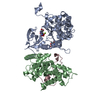 3miaC 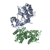 3blhS C: citing same article ( S: Starting model for refinement |
|---|---|
| Similar structure data |
-Links
 PDBj
PDBj


- Assembly
| Deposited unit | 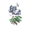
| ||||||||
|---|---|---|---|---|---|---|---|---|---|
| 1 |
| ||||||||
| Unit cell |
|
-Components
| #1: Protein | / Cyclin-dependent kinase 9 / Serine/threonine-protein kinase PITALRE / Cell division cycle 2-like ...Cyclin-dependent kinase 9 / Serine/threonine-protein kinase PITALRE / Cell division cycle 2-like protein kinase 4 / C-2K Mass: 40692.152 Da / Num. of mol.: 1 / Fragment: UNP residues 1-345, Protein kinase domain Source method: isolated from a genetically manipulated source Source: (gene. exp.) Homo sapiens (human) / Gene: CDC2L4, CDK9 / Plasmid: pENTR/SD/D-TOPO vector (Invitrogen K2420) / Cell line (production host): BL21 insect cells / Production host:   Spodoptera frugiperda (fall armyworm) / Strain (production host): SF9 Spodoptera frugiperda (fall armyworm) / Strain (production host): SF9References: UniProt: P50750, cyclin-dependent kinase, [RNA-polymerase]-subunit kinase | ||
|---|---|---|---|
| #2: Protein | Mass: 30877.320 Da / Num. of mol.: 1 / Fragment: UNP residues 1-266 Source method: isolated from a genetically manipulated source Source: (gene. exp.) Homo sapiens (human) / Gene: CCNT1, Cyclin T1 / Plasmid: pENTR/SD/D-TOPO vector (Invitrogen K2420) / Cell line (production host): BL21 insect cells / Production host: Spodoptera frugiperda (fall armyworm) / Strain (production host): SF9 / References: UniProt: O60563 | ||
| #3: Protein | Mass: 9848.327 Da / Num. of mol.: 1 Source method: isolated from a genetically manipulated source Source: (gene. exp.) Human immunodeficiency virus type 1 / Strain: isolate HXB2 group M subtype B / Gene: tat / Plasmid: pENTR/SD/D-TOPO vector (Invitrogen K2420) / Cell line (production host): BL21 insect cells / Production host: Spodoptera frugiperda (fall armyworm) / Strain (production host): SF9 / References: UniProt: P04608 | ||
| #4: Chemical |   Mass: 65.409 Da / Num. of mol.: 2 / Source method: obtained synthetically / Formula: Zn Mass: 65.409 Da / Num. of mol.: 2 / Source method: obtained synthetically / Formula: Zn#5: Water | ChemComp-HOH / | Water Mass: 18.015 Da / Num. of mol.: 341 / Source method: isolated from a natural source / Formula: H2O Mass: 18.015 Da / Num. of mol.: 341 / Source method: isolated from a natural source / Formula: H2O |
-Experimental details
-Experiment
| Experiment | Method: X-RAY DIFFRACTION / Number of used crystals: 1 |
|---|
- Sample preparation
| Crystal | Density Matthews: 2.96 Å3/Da / Density % sol: 58.43 % |
|---|---|
| Crystal grow | Temperature: 295 K / Method: vapor diffusion, sitting drop / pH: 7.5 Details: 50 mM HEPES buffer (pH 7.5), 4.25-5% PEG 20,000, 1 mM TCEP, and 20 mM glycyl-glycyl-glycine , VAPOR DIFFUSION, SITTING DROP, temperature 295.0K |
-Data collection
| Diffraction | Mean temperature: 100 K |
|---|---|
| Diffraction source | Source: SYNCHROTRON / Site: APS  / Beamline: 24-ID-C / Wavelength: 0.9795 Å / Beamline: 24-ID-C / Wavelength: 0.9795 Å |
| Detector | Type: ADSC QUANTUM 315 / Detector: CCD / Date: Oct 17, 2009 |
| Radiation | Protocol: SINGLE WAVELENGTH / Monochromatic (M) / Laue (L): M / Scattering type: x-ray |
| Radiation wavelength | Wavelength: 0.9795 Å / Relative weight: 1 |
| Reflection | Resolution: 2.1→40 Å / Num. obs: 51839 / % possible obs: 90.9 % / Observed criterion σ(F): -2 / Redundancy: 2.5 % / Biso Wilson estimate: 31.7 Å2 / Rmerge(I) obs: 0.055 / Net I/σ(I): 35 |
| Reflection shell | Resolution: 2.1→2.14 Å / Redundancy: 2.2 % / Rmerge(I) obs: 0.52 / Mean I/σ(I) obs: 3.7 / Num. unique all: 2531 / % possible all: 89 |
- Processing
| Software |
| ||||||||||||||||||||||||||||||||||||||||||||||||||||||||||||||||||||||||||||||||
|---|---|---|---|---|---|---|---|---|---|---|---|---|---|---|---|---|---|---|---|---|---|---|---|---|---|---|---|---|---|---|---|---|---|---|---|---|---|---|---|---|---|---|---|---|---|---|---|---|---|---|---|---|---|---|---|---|---|---|---|---|---|---|---|---|---|---|---|---|---|---|---|---|---|---|---|---|---|---|---|---|---|
| Refinement | Method to determine structure: MOLECULAR REPLACEMENT Starting model: PDB ENTRY 3blh Resolution: 2.1→30.63 Å / Rfactor Rfree error: 0.005 / Data cutoff high absF: 2384386.28 / Data cutoff low absF: 0 / Isotropic thermal model: RESTRAINED / Cross valid method: THROUGHOUT / σ(F): 0 / Stereochemistry target values: Engh & Huber
| ||||||||||||||||||||||||||||||||||||||||||||||||||||||||||||||||||||||||||||||||
| Solvent computation | Solvent model: FLAT MODEL / Bsol: 58.0275 Å2 / ksol: 0.36834 e/Å3 | ||||||||||||||||||||||||||||||||||||||||||||||||||||||||||||||||||||||||||||||||
| Displacement parameters | Biso mean: 46.7 Å2
| ||||||||||||||||||||||||||||||||||||||||||||||||||||||||||||||||||||||||||||||||
| Refine analyze |
| ||||||||||||||||||||||||||||||||||||||||||||||||||||||||||||||||||||||||||||||||
| Refinement step | Cycle: LAST / Resolution: 2.1→30.63 Å
| ||||||||||||||||||||||||||||||||||||||||||||||||||||||||||||||||||||||||||||||||
| Refine LS restraints |
| ||||||||||||||||||||||||||||||||||||||||||||||||||||||||||||||||||||||||||||||||
| Refine LS restraints NCS | NCS model details: NONE | ||||||||||||||||||||||||||||||||||||||||||||||||||||||||||||||||||||||||||||||||
| LS refinement shell | Resolution: 2.1→2.23 Å / Rfactor Rfree error: 0.02 / Total num. of bins used: 6
| ||||||||||||||||||||||||||||||||||||||||||||||||||||||||||||||||||||||||||||||||
| Xplor file |
|
