 Movie
Movie Controller

[English] 日本語
 Yorodumi
- PDB-3mez: X-ray structural analysis of a mannose specific lectin from dutch... -
+ Open data

- Basic information
| Entry | Database: PDB / ID: 3mez | ||||||
|---|---|---|---|---|---|---|---|
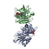
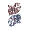
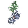
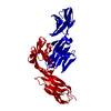
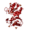
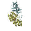
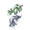
| Title | X-ray structural analysis of a mannose specific lectin from dutch crocus (crocus vernus) | ||||||
 Components Components |
| ||||||
 Keywords Keywords | SUGAR BINDING PROTEIN /  Lectin / Crocus / Heterotetramer Lectin / Crocus / Heterotetramer | ||||||
| Function / homology |  Function and homology information Function and homology information | ||||||
| Biological species |  Crocus vernus (plant) Crocus vernus (plant) | ||||||
| Method | X-RAY DIFFRACTION / SYNCHROTRON / MIR / Resolution: 1.94 Å | ||||||
 Authors Authors | Akrem, A. / Meyer, A. / Perbandt, M. / Voelter, W. / Buck, F. / Betzel, C. | ||||||
 Citation Citation | Journal: To be Published Title: X-ray structural analysis of a mannose specific lectin from dutch crocus (crocus vernus) Authors: Akrem, A. | ||||||
| History |
|
- Structure visualization
| Structure viewer | Molecule: MolmilJmol/JSmol |
|---|
- Downloads & links
-Download
| PDBx/mmCIF format | 3mez.cif.gz | 105.1 KB | Display | PDBx/mmCIF format |
|---|---|---|---|---|
| PDB format | pdb3mez.ent.gz | 81 KB | Display | PDB format |
| PDBx/mmJSON format | 3mez.json.gz | Tree view | PDBx/mmJSON format | |
| Others |  Other downloads Other downloads |
-Validation report
| Arichive directory | https://data.pdbj.org/pub/pdb/validation_reports/me/3mezftp://data.pdbj.org/pub/pdb/validation_reports/me/3mez | HTTPS FTP |
|---|
-Related structure data
| Related structure data | 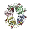 1msaS S: Starting model for refinement |
|---|---|
| Similar structure data |


-Links
 PDBj
PDBj
- Assembly
| Deposited unit | 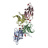
| ||||||||
|---|---|---|---|---|---|---|---|---|---|
| 1 |
| ||||||||
| Unit cell |
|
-Components
| #1: Protein | Mass: 12013.144 Da / Num. of mol.: 2 / Source method: isolated from a natural source / Details: Bulb / Source: (natural) Crocus vernus (plant) / References: UniProt: P86626#2: Protein | Mass: 12478.987 Da / Num. of mol.: 2 / Source method: isolated from a natural source / Details: Bulb / Source: (natural) Crocus vernus (plant) / References: UniProt: P86626#3: Chemical | Glycerol  Mass: 92.094 Da / Num. of mol.: 3 / Source method: obtained synthetically / Formula: C3H8O3 Mass: 92.094 Da / Num. of mol.: 3 / Source method: obtained synthetically / Formula: C3H8O3#4: Chemical | ChemComp-FMT / Formic acid  Mass: 46.025 Da / Num. of mol.: 8 / Source method: obtained synthetically / Formula: CH2O2 Mass: 46.025 Da / Num. of mol.: 8 / Source method: obtained synthetically / Formula: CH2O2#5: Water | ChemComp-HOH / | Water Mass: 18.015 Da / Num. of mol.: 295 / Source method: isolated from a natural source / Formula: H2O Mass: 18.015 Da / Num. of mol.: 295 / Source method: isolated from a natural source / Formula: H2OSequence details | AUTHOR WILL CONTACT UNIPROT FOR MODIFICATI | |
|---|
-Experimental details
-Experiment
| Experiment | Method: X-RAY DIFFRACTION / Number of used crystals: 1 |
|---|
- Sample preparation
| Crystal | Density Matthews: 2.57 Å3/Da / Density % sol: 52.08 % |
|---|---|
| Crystal grow | Temperature: 293 K / Method: vapor diffusion, hanging drop / pH: 7.5 Details: 4M Sodium formate, pH 7.5, VAPOR DIFFUSION, HANGING DROP, temperature 293K |
-Data collection
| Diffraction | Mean temperature: 77 K |
|---|---|
| Diffraction source | Source: SYNCHROTRON / Site: EMBL/DESY, HAMBURG  / Beamline: X13 / Wavelength: 0.8123 Å / Beamline: X13 / Wavelength: 0.8123 Å |
| Detector | Type: MAR CCD 165 mm / Detector: CCD / Date: May 16, 2009 |
| Radiation | Protocol: SINGLE WAVELENGTH / Monochromatic (M) / Laue (L): M / Scattering type: x-ray |
| Radiation wavelength | Wavelength: 0.8123 Å / Relative weight: 1 |
| Reflection | Resolution: 1.94→30 Å / Num. obs: 48106 / Observed criterion σ(I): 3.84 / Redundancy: 4.3 % / Rmerge(I) obs: 0.098 |
| Reflection shell | Resolution: 1.94→2 Å / Redundancy: 4.3 % / Rmerge(I) obs: 0.33 |
- Processing
| Software |
| ||||||||||||||||||||||||||||||||||||||||||||||||||||||||||||||||||||||||||||||||||||||||||||||||||||||||||||||||||||||||||||||||||||||||||||||||||||||||||||||||||||||||||
|---|---|---|---|---|---|---|---|---|---|---|---|---|---|---|---|---|---|---|---|---|---|---|---|---|---|---|---|---|---|---|---|---|---|---|---|---|---|---|---|---|---|---|---|---|---|---|---|---|---|---|---|---|---|---|---|---|---|---|---|---|---|---|---|---|---|---|---|---|---|---|---|---|---|---|---|---|---|---|---|---|---|---|---|---|---|---|---|---|---|---|---|---|---|---|---|---|---|---|---|---|---|---|---|---|---|---|---|---|---|---|---|---|---|---|---|---|---|---|---|---|---|---|---|---|---|---|---|---|---|---|---|---|---|---|---|---|---|---|---|---|---|---|---|---|---|---|---|---|---|---|---|---|---|---|---|---|---|---|---|---|---|---|---|---|---|---|---|---|---|---|---|
| Refinement | Method to determine structure: MIR Starting model: PDB ENTRY 1MSA Resolution: 1.94→30 Å / Cor.coef. Fo:Fc: 0.959 / Cor.coef. Fo:Fc free: 0.934 / SU B: 3.405 / SU ML: 0.098 / Isotropic thermal model: Isotropic / Cross valid method: THROUGHOUT / ESU R: 0.18 / ESU R Free: 0.163 / Stereochemistry target values: MAXIMUM LIKELIHOOD / Details: HYDROGENS HAVE BEEN ADDED IN THE RIDING POSITIONS
| ||||||||||||||||||||||||||||||||||||||||||||||||||||||||||||||||||||||||||||||||||||||||||||||||||||||||||||||||||||||||||||||||||||||||||||||||||||||||||||||||||||||||||
| Solvent computation | Ion probe radii: 0.8 Å / Shrinkage radii: 0.8 Å / VDW probe radii: 1.4 Å / Solvent model: MASK | ||||||||||||||||||||||||||||||||||||||||||||||||||||||||||||||||||||||||||||||||||||||||||||||||||||||||||||||||||||||||||||||||||||||||||||||||||||||||||||||||||||||||||
| Displacement parameters | Biso mean: 25.909 Å2
| ||||||||||||||||||||||||||||||||||||||||||||||||||||||||||||||||||||||||||||||||||||||||||||||||||||||||||||||||||||||||||||||||||||||||||||||||||||||||||||||||||||||||||
| Refinement step | Cycle: LAST / Resolution: 1.94→30 Å
| ||||||||||||||||||||||||||||||||||||||||||||||||||||||||||||||||||||||||||||||||||||||||||||||||||||||||||||||||||||||||||||||||||||||||||||||||||||||||||||||||||||||||||
| Refine LS restraints |
| ||||||||||||||||||||||||||||||||||||||||||||||||||||||||||||||||||||||||||||||||||||||||||||||||||||||||||||||||||||||||||||||||||||||||||||||||||||||||||||||||||||||||||
| LS refinement shell | Resolution: 1.94→1.99 Å / Total num. of bins used: 20
|
