 Movie
Movie Controller

[English] 日本語
 Yorodumi
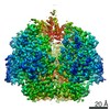
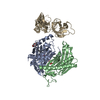
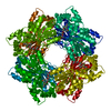
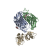
- PDB-3m85: Archaeoglobus fulgidus exosome y70a with RNA bound to the active site -
+ Open data

- Basic information
| Entry | Database: PDB / ID: 3m85 | ||||||
|---|---|---|---|---|---|---|---|
| Title | Archaeoglobus fulgidus exosome y70a with RNA bound to the active site | ||||||
 Components Components |
| ||||||
 Keywords Keywords | HYDROLASE/RNA / exosome /  RNA / Exonuclease / Hydrolase / Nuclease / HYDROLASE-RNA complex RNA / Exonuclease / Hydrolase / Nuclease / HYDROLASE-RNA complex | ||||||
| Function / homology |  Function and homology information Function and homology informationexosome (RNase complex) / RNA catabolic process / RNA processing / Hydrolases; Acting on ester bonds; Exoribonucleases producing 5'-phosphomonoesters / 3'-5'-RNA exonuclease activity / RNA binding / zinc ion binding / cytoplasmSimilarity search - Function | ||||||
| Biological species |   Archaeoglobus fulgidus (archaea)archaeoglobus fulgidus (archaea) Archaeoglobus fulgidus (archaea)archaeoglobus fulgidus (archaea) | ||||||
| Method | X-RAY DIFFRACTION / SYNCHROTRON / MOLECULAR REPLACEMENT / Resolution: 3 Å | ||||||
 Authors Authors | Hartung, S. / Hopfner, K.-P. | ||||||
 Citation Citation | Journal: Nucleic Acids Res. / Year: 2010 Title: Quantitative analysis of processive RNA degradation by the archaeal RNA exosome Authors: Hartung, S. / Niederberger, T. / Hartung, M. / Tresch, A. / Hopfner, K.-P. | ||||||
| History |
|
- Structure visualization
| Structure viewer | Molecule: MolmilJmol/JSmol |
|---|
- Downloads & links
-Download
| PDBx/mmCIF format | 3m85.cif.gz | 406.5 KB | Display | PDBx/mmCIF format |
|---|---|---|---|---|
| PDB format | pdb3m85.ent.gz | 330.1 KB | Display | PDB format |
| PDBx/mmJSON format | 3m85.json.gz | Tree view | PDBx/mmJSON format | |
| Others |  Other downloads Other downloads |
-Validation report
| Arichive directory | https://data.pdbj.org/pub/pdb/validation_reports/m8/3m85ftp://data.pdbj.org/pub/pdb/validation_reports/m8/3m85 | HTTPS FTP |
|---|
-Related structure data
| Related structure data |  3m7nC  2ab1S C: citing same article ( S: Starting model for refinement |
|---|---|
| Similar structure data |


-Links
 PDBj
PDBj


- Assembly
| Deposited unit | 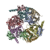
| ||||||||
|---|---|---|---|---|---|---|---|---|---|
| 1 |
| ||||||||
| Unit cell |
| ||||||||
| Components on special symmetry positions |
|
-Components
-Probable exosome complex exonuclease ... , 2 types, 6 molecules DEFGHI
| #2: Protein | Mass: 28860.266 Da / Num. of mol.: 3 / Mutation: R65E Source method: isolated from a genetically manipulated source Source: (gene. exp.) archaeoglobus fulgidus (archaea) / Gene: AF_0493 / Plasmid: pET-21 / Production host:  Escherichia coli (E. coli) / Strain (production host): Rosetta DE3 Escherichia coli (E. coli) / Strain (production host): Rosetta DE3References: UniProt: O29757, Hydrolases; Acting on ester bonds; Exoribonucleases producing 5'-phosphomonoesters#3: Protein | Mass: 28585.512 Da / Num. of mol.: 3 / Mutation: Y70A Source method: isolated from a genetically manipulated source Source: (gene. exp.) archaeoglobus fulgidus (archaea) / Gene: AF_0494 / Plasmid: pET-21 / Production host: Escherichia coli (E. coli) / Strain (production host): Rosetta DE3References: UniProt: O29756, Hydrolases; Acting on ester bonds; Exoribonucleases producing 5'-phosphomonoesters |
|---|
-Protein / RNA chain , 2 types, 6 molecules ABCXYZ
| #1: Protein | Mass: 19625.604 Da / Num. of mol.: 3 Source method: isolated from a genetically manipulated source Source: (gene. exp.) Archaeoglobus fulgidus (archaea) / Gene: AF_0206 / Plasmid: pET-28 / Production host: Escherichia coli (E. coli) / Strain (production host): Rosetta DE3 / References: UniProt: O30033#4: RNA chain | Mass: 1787.117 Da / Num. of mol.: 3 / Source method: obtained synthetically |
|---|
-Non-polymers , 2 types, 9 molecules 

| #5: Chemical |  Mass: 65.409 Da / Num. of mol.: 3 / Source method: obtained synthetically / Formula: Zn Mass: 65.409 Da / Num. of mol.: 3 / Source method: obtained synthetically / Formula: Zn#6: Water | ChemComp-HOH / | WaterMass: 18.015 Da / Num. of mol.: 6 / Source method: isolated from a natural source / Formula: H2O |
|---|
-Experimental details
-Experiment
| Experiment | Method: X-RAY DIFFRACTION / Number of used crystals: 1 |
|---|
- Sample preparation
| Crystal | Density Matthews: 2.64 Å3/Da / Density % sol: 53.4 % |
|---|---|
| Crystal grow | Temperature: 293 K / Method: vapor diffusion, sitting drop / pH: 4.6 Details: 0.1M Na Acetate, pH 4.6, 30% MPD, 100mM NaCl, VAPOR DIFFUSION, SITTING DROP, temperature 293K |
-Data collection
| Diffraction | Mean temperature: 100 K |
|---|---|
| Diffraction source | Source: SYNCHROTRON / Site: SLS  / Beamline: X06SA / Wavelength: 1.006 Å / Beamline: X06SA / Wavelength: 1.006 Å |
| Detector | Type: MARMOSAIC 225 mm CCD / Detector: CCD / Date: Sep 6, 2006 |
| Radiation | Monochromator: LN2 cooled fixed-exit Si(111) monochromator / Protocol: SINGLE WAVELENGTH / Monochromatic (M) / Laue (L): M / Scattering type: x-ray |
| Radiation wavelength | Wavelength: 1.006 Å / Relative weight: 1 |
| Reflection | Resolution: 3→50 Å / Num. all: 51207 / Num. obs: 51203 / % possible obs: 98.9 % / Observed criterion σ(F): 2 / Observed criterion σ(I): 2 |
| Reflection shell | Resolution: 3→3.1 Å / Redundancy: 7.22 % / Num. unique all: 50904 / % possible all: 98.9 |
- Processing
| Software |
| |||||||||||||||||||||||||||||||||||||||||||||||||||||||||||||||||||||||||||||||||||||||||||||||||||||||||||||||||||||||||||||||||||||||||||||||||||||||||||||||||||||||||||||||||||||||||||||||||||||||||||||||||||||||||
|---|---|---|---|---|---|---|---|---|---|---|---|---|---|---|---|---|---|---|---|---|---|---|---|---|---|---|---|---|---|---|---|---|---|---|---|---|---|---|---|---|---|---|---|---|---|---|---|---|---|---|---|---|---|---|---|---|---|---|---|---|---|---|---|---|---|---|---|---|---|---|---|---|---|---|---|---|---|---|---|---|---|---|---|---|---|---|---|---|---|---|---|---|---|---|---|---|---|---|---|---|---|---|---|---|---|---|---|---|---|---|---|---|---|---|---|---|---|---|---|---|---|---|---|---|---|---|---|---|---|---|---|---|---|---|---|---|---|---|---|---|---|---|---|---|---|---|---|---|---|---|---|---|---|---|---|---|---|---|---|---|---|---|---|---|---|---|---|---|---|---|---|---|---|---|---|---|---|---|---|---|---|---|---|---|---|---|---|---|---|---|---|---|---|---|---|---|---|---|---|---|---|---|---|---|---|---|---|---|---|---|---|---|---|---|---|---|---|---|
| Refinement | Method to determine structure: MOLECULAR REPLACEMENT Starting model: PDB ENTRY 2AB1 Resolution: 3→19.984 Å / SU ML: 0.38 / σ(F): 1.39 / Stereochemistry target values: ML
| |||||||||||||||||||||||||||||||||||||||||||||||||||||||||||||||||||||||||||||||||||||||||||||||||||||||||||||||||||||||||||||||||||||||||||||||||||||||||||||||||||||||||||||||||||||||||||||||||||||||||||||||||||||||||
| Solvent computation | Shrinkage radii: 0.9 Å / VDW probe radii: 1.11 Å / Solvent model: FLAT BULK SOLVENT MODEL / Bsol: 43.417 Å2 / ksol: 0.28 e/Å3 | |||||||||||||||||||||||||||||||||||||||||||||||||||||||||||||||||||||||||||||||||||||||||||||||||||||||||||||||||||||||||||||||||||||||||||||||||||||||||||||||||||||||||||||||||||||||||||||||||||||||||||||||||||||||||
| Refine analyze | Luzzati coordinate error obs: 0.38 Å | |||||||||||||||||||||||||||||||||||||||||||||||||||||||||||||||||||||||||||||||||||||||||||||||||||||||||||||||||||||||||||||||||||||||||||||||||||||||||||||||||||||||||||||||||||||||||||||||||||||||||||||||||||||||||
| Refinement step | Cycle: LAST / Resolution: 3→19.984 Å
| |||||||||||||||||||||||||||||||||||||||||||||||||||||||||||||||||||||||||||||||||||||||||||||||||||||||||||||||||||||||||||||||||||||||||||||||||||||||||||||||||||||||||||||||||||||||||||||||||||||||||||||||||||||||||
| Refine LS restraints |
| |||||||||||||||||||||||||||||||||||||||||||||||||||||||||||||||||||||||||||||||||||||||||||||||||||||||||||||||||||||||||||||||||||||||||||||||||||||||||||||||||||||||||||||||||||||||||||||||||||||||||||||||||||||||||
| LS refinement shell |
|
