 Movie
Movie Controller

[English] 日本語
 Yorodumi
- PDB-3m6k: Crystal Structure of N-terminal 44 kDa fragment of topoisomerase ... -
+ Open data

- Basic information
| Entry | Database: PDB / ID: 3m6k | ||||||
|---|---|---|---|---|---|---|---|
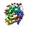
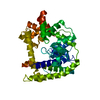
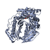
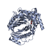
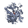
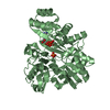
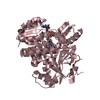
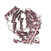
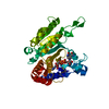
| Title | Crystal Structure of N-terminal 44 kDa fragment of topoisomerase V in the presence of guanidium hydrochloride | ||||||
 Components Components | Topoisomerase V | ||||||
 Keywords Keywords | ISOMERASE / helix-hairpin-helix / topoisomerase / conformational change in protein / guanidium hydrochloride | ||||||
| Function / homology |  Function and homology information Function and homology information | ||||||
| Biological species |   Methanopyrus kandleri (archaea) Methanopyrus kandleri (archaea) | ||||||
| Method | X-RAY DIFFRACTION / SYNCHROTRON / SAD / Resolution: 2.6 Å | ||||||
 Authors Authors | Rajan, R. / Taneja, B. / Mondragon, A. | ||||||
 Citation Citation | Journal: Structure / Year: 2010 Title: Structures of minimal catalytic fragments of topoisomerase v reveals conformational changes relevant for DNA binding. Authors: Rajan, R. / Taneja, B. / Mondragon, A. | ||||||
| History |
|
- Structure visualization
| Structure viewer | Molecule: MolmilJmol/JSmol |
|---|
- Downloads & links
-Download
| PDBx/mmCIF format | 3m6k.cif.gz | 300.6 KB | Display | PDBx/mmCIF format |
|---|---|---|---|---|
| PDB format | pdb3m6k.ent.gz | 257.9 KB | Display | PDB format |
| PDBx/mmJSON format | 3m6k.json.gz | Tree view | PDBx/mmJSON format | |
| Others |  Other downloads Other downloads |
-Validation report
| Arichive directory | https://data.pdbj.org/pub/pdb/validation_reports/m6/3m6kftp://data.pdbj.org/pub/pdb/validation_reports/m6/3m6k | HTTPS FTP |
|---|
-Related structure data


-Links
 PDBj
PDBj


- Assembly
| Deposited unit | 
| ||||||||||||||||||||||||||||||||||||||||||||||||
|---|---|---|---|---|---|---|---|---|---|---|---|---|---|---|---|---|---|---|---|---|---|---|---|---|---|---|---|---|---|---|---|---|---|---|---|---|---|---|---|---|---|---|---|---|---|---|---|---|---|
| 1 | 
| ||||||||||||||||||||||||||||||||||||||||||||||||
| 2 | 
| ||||||||||||||||||||||||||||||||||||||||||||||||
| Unit cell |
| ||||||||||||||||||||||||||||||||||||||||||||||||
| Components on special symmetry positions |
| ||||||||||||||||||||||||||||||||||||||||||||||||
| Noncrystallographic symmetry (NCS) | NCS domain:
NCS domain segments:
NCS ensembles :
|
-Components
| #1: Protein | Mass: 44232.012 Da / Num. of mol.: 2 / Fragment: N-terminal 44 kDa fragment (Topo-44) Source method: isolated from a genetically manipulated source Source: (gene. exp.) Methanopyrus kandleri (archaea) / Strain: AV19 / Gene: MK1436, top5, Topoisomerase V / Plasmid: pET15b / Production host:  Escherichia coli (E. coli) / Strain (production host): BL21 Rosetta (DE3) / References: UniProt: Q977W1 Escherichia coli (E. coli) / Strain (production host): BL21 Rosetta (DE3) / References: UniProt: Q977W1#2: Chemical | Phosphate  Mass: 94.971 Da / Num. of mol.: 3 / Source method: obtained synthetically / Formula: PO4 Mass: 94.971 Da / Num. of mol.: 3 / Source method: obtained synthetically / Formula: PO4#3: Water | ChemComp-HOH / | Water Mass: 18.015 Da / Num. of mol.: 30 / Source method: isolated from a natural source / Formula: H2O Mass: 18.015 Da / Num. of mol.: 30 / Source method: isolated from a natural source / Formula: H2O |
|---|
-Experimental details
-Experiment
| Experiment | Method: X-RAY DIFFRACTION / Number of used crystals: 1 |
|---|
- Sample preparation
| Crystal | Density Matthews: 2.43 Å3/Da / Density % sol: 49.36 % |
|---|---|
| Crystal grow | Temperature: 303 K / Method: vapor diffusion, hanging drop / pH: 5 Details: 0.1 M phosphate-citrate pH 5, 0.2 M NaCl, 16% PEG 8000, 1M Guanidium hydrochloride, VAPOR DIFFUSION, HANGING DROP, temperature 303K |
-Data collection
| Diffraction |
| ||||||||||||||||||
|---|---|---|---|---|---|---|---|---|---|---|---|---|---|---|---|---|---|---|---|
| Diffraction source |
| ||||||||||||||||||
| Detector |
| ||||||||||||||||||
| Radiation |
| ||||||||||||||||||
| Radiation wavelength |
| ||||||||||||||||||
| Reflection | Resolution: 2.6→29.5 Å / Num. all: 28160 / Num. obs: 28151 / % possible obs: 99.9 % / Redundancy: 8.1 % / Biso Wilson estimate: 42.1 Å2 / Rmerge(I) obs: 0.079 / Rsym value: 0.074 / Net I/σ(I): 19 | ||||||||||||||||||
| Reflection shell | Resolution: 2.6→2.72 Å / Redundancy: 6 % / Rmerge(I) obs: 0.572 / Mean I/σ(I) obs: 3.2 / Num. unique all: 3331 / Rsym value: 0.522 / % possible all: 100 |

- Processing
| Software |
| |||||||||||||||||||||||||||||||||||||||||||||||||||||||||||||||||||||||||||||||||||||||||||||||||||||||||||||||||||||||||||||
|---|---|---|---|---|---|---|---|---|---|---|---|---|---|---|---|---|---|---|---|---|---|---|---|---|---|---|---|---|---|---|---|---|---|---|---|---|---|---|---|---|---|---|---|---|---|---|---|---|---|---|---|---|---|---|---|---|---|---|---|---|---|---|---|---|---|---|---|---|---|---|---|---|---|---|---|---|---|---|---|---|---|---|---|---|---|---|---|---|---|---|---|---|---|---|---|---|---|---|---|---|---|---|---|---|---|---|---|---|---|---|---|---|---|---|---|---|---|---|---|---|---|---|---|---|---|---|
| Refinement | Method to determine structure: SAD / Resolution: 2.6→29.14 Å / Cor.coef. Fo:Fc: 0.928 / Cor.coef. Fo:Fc free: 0.898 / SU B: 28.292 / SU ML: 0.279 / Cross valid method: THROUGHOUT / ESU R: 0.963 / ESU R Free: 0.363 / Stereochemistry target values: MAXIMUM LIKELIHOOD / Details: HYDROGENS HAVE BEEN ADDED IN THE RIDING POSITIONS
| |||||||||||||||||||||||||||||||||||||||||||||||||||||||||||||||||||||||||||||||||||||||||||||||||||||||||||||||||||||||||||||
| Solvent computation | Ion probe radii: 0.8 Å / Shrinkage radii: 0.8 Å / VDW probe radii: 1.2 Å / Solvent model: MASK | |||||||||||||||||||||||||||||||||||||||||||||||||||||||||||||||||||||||||||||||||||||||||||||||||||||||||||||||||||||||||||||
| Displacement parameters | Biso mean: 55.918 Å2
| |||||||||||||||||||||||||||||||||||||||||||||||||||||||||||||||||||||||||||||||||||||||||||||||||||||||||||||||||||||||||||||
| Refinement step | Cycle: LAST / Resolution: 2.6→29.14 Å
| |||||||||||||||||||||||||||||||||||||||||||||||||||||||||||||||||||||||||||||||||||||||||||||||||||||||||||||||||||||||||||||
| Refine LS restraints |
| |||||||||||||||||||||||||||||||||||||||||||||||||||||||||||||||||||||||||||||||||||||||||||||||||||||||||||||||||||||||||||||
| Refine LS restraints NCS | Dom-ID: 1 / Auth asym-ID: B / Refine-ID: X-RAY DIFFRACTION
| |||||||||||||||||||||||||||||||||||||||||||||||||||||||||||||||||||||||||||||||||||||||||||||||||||||||||||||||||||||||||||||
| LS refinement shell | Resolution: 2.6→2.667 Å / Total num. of bins used: 20
| |||||||||||||||||||||||||||||||||||||||||||||||||||||||||||||||||||||||||||||||||||||||||||||||||||||||||||||||||||||||||||||
| Refinement TLS params. | Method: refined / Refine-ID: X-RAY DIFFRACTION
| |||||||||||||||||||||||||||||||||||||||||||||||||||||||||||||||||||||||||||||||||||||||||||||||||||||||||||||||||||||||||||||
| Refinement TLS group |
|
