Mass: 18.015 Da / Num. of mol.: 333 / Source method: isolated from a natural source / Formula: H2O
-
Experimental details
-
Experiment
Experiment
Method: X-RAY DIFFRACTION / Number of used crystals: 1
-
Sample preparation
Crystal
Density Matthews: 1.85 Å3/Da / Density % sol: 33.56 %
Crystal grow
Temperature: 298 K / Method: vapor diffusion, hanging drop / pH: 7.5 Details: LIGAND-FREE PROTEIN CRYSTAL GROWN IN SOLUTION CONTAINING 30% PEG 400, 0.1 M HEPES pH7.5, 0.2 M CaCl2, THEN SOAKED IN MOTHER LIQUOR CONTAINING 0.2M MNCL2 AND 20 mM INHIBITOR FOR 15 min, VAPOR ...Details: LIGAND-FREE PROTEIN CRYSTAL GROWN IN SOLUTION CONTAINING 30% PEG 400, 0.1 M HEPES pH7.5, 0.2 M CaCl2, THEN SOAKED IN MOTHER LIQUOR CONTAINING 0.2M MNCL2 AND 20 mM INHIBITOR FOR 15 min, VAPOR DIFFUSION, HANGING DROP, temperature 298K
Resolution: 1.55→19.92 Å / Occupancy max: 1 / Occupancy min: 0 / SU ML: 0.18 / Isotropic thermal model: Isotropic / σ(F): 1.48 / Phase error: 19.7 / Stereochemistry target values: ML Details: ANISOTROPIC B VALUES DERIVED FROM FROM TLS REFINEMENT, HYDROGENS ARE INCLUDED AS RIDING MODEL
Rfactor
Num. reflection
% reflection
Rfree
0.201
1875
5.12 %
Rwork
0.175
-
-
obs
0.176
36608
99.9 %
Solvent computation
Shrinkage radii: 0.9 Å / VDW probe radii: 1.11 Å / Solvent model: FLAT BULK SOLVENT MODEL / Bsol: 81.42 Å2 / ksol: 0.44 e/Å3
Displacement parameters
Biso mean: 22.1 Å2
Baniso -1
Baniso -2
Baniso -3
1-
0.145 Å2
0 Å2
-0 Å2
2-
-
0.145 Å2
0 Å2
3-
-
-
-0.289 Å2
Refinement step
Cycle: LAST / Resolution: 1.55→19.92 Å
Protein
Nucleic acid
Ligand
Solvent
Total
Num. atoms
2250
0
14
333
2597
Refine LS restraints
Refine-ID
Type
Dev ideal
Number
X-RAY DIFFRACTION
f_bond_d
0.006
4565
X-RAY DIFFRACTION
f_angle_d
0.939
8253
X-RAY DIFFRACTION
f_dihedral_angle_d
16.338
1174
X-RAY DIFFRACTION
f_chiral_restr
0.073
353
X-RAY DIFFRACTION
f_plane_restr
0.004
705
LS refinement shell
Resolution (Å)
Rfactor Rfree
Num. reflection Rfree
Rfactor Rwork
Num. reflection Rwork
Refine-ID
% reflection obs (%)
1.55-1.5919
0.3595
149
0.3309
2614
X-RAY DIFFRACTION
100
1.5919-1.6387
0.3473
148
0.299
2603
X-RAY DIFFRACTION
100
1.6387-1.6916
0.2909
142
0.2868
2630
X-RAY DIFFRACTION
100
1.6916-1.752
0.3136
130
0.2564
2644
X-RAY DIFFRACTION
100
1.752-1.8221
0.2941
147
0.2295
2634
X-RAY DIFFRACTION
100
1.8221-1.905
0.2299
148
0.1879
2625
X-RAY DIFFRACTION
100
1.905-2.0053
0.2106
131
0.1573
2667
X-RAY DIFFRACTION
100
2.0053-2.1308
0.1615
135
0.143
2656
X-RAY DIFFRACTION
100
2.1308-2.2951
0.1675
146
0.1317
2672
X-RAY DIFFRACTION
100
2.2951-2.5257
0.1745
145
0.1372
2677
X-RAY DIFFRACTION
100
2.5257-2.8902
0.142
148
0.1324
2699
X-RAY DIFFRACTION
100
2.8902-3.6377
0.1587
138
0.131
2750
X-RAY DIFFRACTION
100
3.6377-19.9245
0.1688
168
0.1669
2862
X-RAY DIFFRACTION
100
Refinement TLS params.
Method: refined / Refine-ID: X-RAY DIFFRACTION
ID
L11 (°2)
L12 (°2)
L13 (°2)
L22 (°2)
L23 (°2)
L33 (°2)
S11 (Å °)
S12 (Å °)
S13 (Å °)
S21 (Å °)
S22 (Å °)
S23 (Å °)
S31 (Å °)
S32 (Å °)
S33 (Å °)
T11 (Å2)
T12 (Å2)
T13 (Å2)
T22 (Å2)
T23 (Å2)
T33 (Å2)
Origin x (Å)
Origin y (Å)
Origin z (Å)
1
0.3391
0.0223
0.1131
0.598
-0.2674
0.4519
-0.0096
0.0112
-0.0016
0.0899
-0.0195
-0.0798
-0.0249
0.0638
-0.0001
0.0677
0.0009
-0.017
0.0588
0.0107
0.0825
18.3371
2.3215
-26.0254
2
0.0161
0.0134
0.0909
0.1442
-0.0843
0.1139
-0.0228
0.0178
0.0308
0.0772
0.0021
-0.0662
-0.0418
0.0084
-0.0051
0.0851
0.0041
-0.0219
0.06
0.0027
0.0734
19.1104
0.7923
-25.526
Refinement TLS group
ID
Refine-ID
Refine TLS-ID
Selection details
1
X-RAY DIFFRACTION
1
CHAIN A AND RESIDUE 68-364
2
X-RAY DIFFRACTION
2
NOT (CHAIN A AND RESIDUE 68-364)
+
About Yorodumi
-
News
-
Feb 9, 2022. New format data for meta-information of EMDB entries
New format data for meta-information of EMDB entries
Version 3 of the EMDB header file is now the official format.
The previous official version 1.9 will be removed from the archive.
In the structure databanks used in Yorodumi, some data are registered as the other names, "COVID-19 virus" and "2019-nCoV". Here are the details of the virus and the list of structure data.
Jan 31, 2019. EMDB accession codes are about to change! (news from PDBe EMDB page)
EMDB accession codes are about to change! (news from PDBe EMDB page)
The allocation of 4 digits for EMDB accession codes will soon come to an end. Whilst these codes will remain in use, new EMDB accession codes will include an additional digit and will expand incrementally as the available range of codes is exhausted. The current 4-digit format prefixed with “EMD-” (i.e. EMD-XXXX) will advance to a 5-digit format (i.e. EMD-XXXXX), and so on. It is currently estimated that the 4-digit codes will be depleted around Spring 2019, at which point the 5-digit format will come into force.
The EM Navigator/Yorodumi systems omit the EMD- prefix.
Related info.:Q: What is EMD? / ID/Accession-code notation in Yorodumi/EM Navigator
Yorodumi is a browser for structure data from EMDB, PDB, SASBDB, etc.
This page is also the successor to EM Navigator detail page, and also detail information page/front-end page for Omokage search.
The word "yorodu" (or yorozu) is an old Japanese word meaning "ten thousand". "mi" (miru) is to see.
Related info.:EMDB / PDB / SASBDB / Comparison of 3 databanks / Yorodumi Search / Aug 31, 2016. New EM Navigator & Yorodumi / Yorodumi Papers / Jmol/JSmol / Function and homology information / Changes in new EM Navigator and Yorodumi
 Movie
Movie Controller

 Yorodumi
 Open data
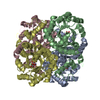
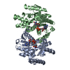
 Basic information
 Components
 Keywords
 HYDROLASE / (alpha/beta)8 barrel
 Function and homology information
 Authors
 Citation
 Structure visualization
 Downloads & links
 Other downloads


 PDBj
PDBj Assembly


 Mass: 186.068 Da / Num. of mol.: 1 / Source method: obtained synthetically / Formula: C4H4F2O6
 Mass: 54.938 Da / Num. of mol.: 1 / Source method: obtained synthetically / Formula: Mn
 Mass: 40.078 Da / Num. of mol.: 1 / Source method: obtained synthetically / Formula: Ca
 Mass: 18.015 Da / Num. of mol.: 333 / Source method: isolated from a natural source / Formula: H2O
 Sample preparation
 Processing