- PDB-3lul: Crystal structure of Putative 4-amino-4-deoxychorismate lyase. (Y... -
+
Open data
ID or keywords:
Loading...
-
Basic information
Entry
Database: PDB / ID: 3lul
Title
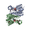
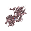
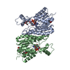
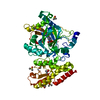
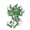
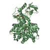
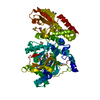
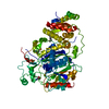
Crystal structure of Putative 4-amino-4-deoxychorismate lyase. (YP_094631.1) from Legionella pneumophila subsp. pneumophila str. Philadelphia 1 at 1.78 A resolution
Components
4-amino-4-deoxychorismate lyase
Keywords
LYASE / Putative 4-amino-4-deoxychorismate lyase. / Structural Genomics / Joint Center for Structural Genomics / JCSG / Protein Structure Initiative / PSI-2 / Pyridoxal phosphate
Mass: 18.015 Da / Num. of mol.: 532 / Source method: isolated from a natural source / Formula: H2O
Sequence details
THE CONSTRUCT WAS EXPRESSED WITH A PURIFICATION TAG MGSDKIHHHHHHENLYFQG. THE TAG WAS REMOVED WITH ...THE CONSTRUCT WAS EXPRESSED WITH A PURIFICATION TAG MGSDKIHHHHHHENLYFQG. THE TAG WAS REMOVED WITH TEV PROTEASE LEAVING ONLY A GLYCINE (0) FOLLOWED BY THE TARGET SEQUENCE.
-
Experimental details
-
Experiment
Experiment
Method: X-RAY DIFFRACTION / Number of used crystals: 1
-
Sample preparation
Crystal
Density Matthews: 2.16 Å3/Da / Density % sol: 42.97 %
Crystal grow
Temperature: 277 K / Method: vapor diffusion, sitting drop / pH: 7.9 Details: 0.2000M (NH4)2HPO4, 20.0000% PEG-3350, No Buffer pH 7.9, NANODROP, VAPOR DIFFUSION, SITTING DROP, temperature 277K
Resolution: 1.78→45.592 Å / Num. obs: 52320 / % possible obs: 99.1 % / Observed criterion σ(I): -3 / Biso Wilson estimate: 19.286 Å2 / Rmerge(I) obs: 0.074 / Net I/σ(I): 11.81
Reflection shell
Resolution (Å)
Rmerge(I) obs
Mean I/σ(I) obs
Num. measured obs
Num. unique obs
Diffraction-ID
% possible all
1.78-1.84
0.538
2.4
17821
4903
1
99.6
1.84-1.92
0.384
3.3
20567
5616
1
99.9
1.92-2
0.264
4.9
17571
4784
1
99.8
2-2.11
0.198
6.4
20009
5471
1
99.9
2.11-2.24
0.144
8.6
18916
5168
1
99.7
2.24-2.41
0.115
10.4
18751
5137
1
99.5
2.41-2.66
0.091
12.6
19756
5414
1
99.4
2.66-3.04
0.064
16.8
18819
5172
1
98.9
3.04-3.83
0.044
23.5
19024
5265
1
98
3.83-45.592
0.035
28.3
19326
5402
1
96.5
-
Phasing
Phasing
Method: MAD
-
Processing
Software
Name
Version
Classification
NB
REFMAC
5.5.0102
refinement
PHENIX
refinement
SHELX
phasing
MolProbity
3beta29
modelbuilding
XSCALE
datascaling
PDB_EXTRACT
3.006
dataextraction
XDS
datareduction
SHELXD
phasing
autoSHARP
phasing
Refinement
Method to determine structure: MAD / Resolution: 1.78→45.592 Å / Cor.coef. Fo:Fc: 0.963 / Cor.coef. Fo:Fc free: 0.941 / Occupancy max: 1 / Occupancy min: 0.23 / SU B: 5.271 / SU ML: 0.074 / TLS residual ADP flag: LIKELY RESIDUAL / Cross valid method: THROUGHOUT / σ(F): 0 / ESU R: 0.121 / ESU R Free: 0.116 Stereochemistry target values: MAXIMUM LIKELIHOOD WITH PHASES Details: 1. HYDROGENS HAVE BEEN ADDED IN THE RIDING POSITIONS. 2. A MET-INHIBITION PROTOCOL WAS USED FOR SELENOMETHIONINE INCORPORATION DURING PROTEIN EXPRESSION. THE OCCUPANCY OF THE SE ATOMS IN THE ...Details: 1. HYDROGENS HAVE BEEN ADDED IN THE RIDING POSITIONS. 2. A MET-INHIBITION PROTOCOL WAS USED FOR SELENOMETHIONINE INCORPORATION DURING PROTEIN EXPRESSION. THE OCCUPANCY OF THE SE ATOMS IN THE MSE RESIDUES WAS REDUCED TO 0.75 FOR THE REDUCED SCATTERING POWER DUE TO PARTIAL S-MET INCORPORATION. 3. ATOM RECORDS CONTAIN RESIDUAL B FACTORS ONLY. 4. ETHYLENE GLYCOL (EDO) AND PHOSPHATE (PO4) MODELED WERE PRESENT IN CRYO/CRYSTALLIZATION BUFFER. 5. ELECTRON DENSITY REVEALS THAT RESIDUE LYS-140 IS COVALENTLY ATTACHED TO PYRIDOXAL-5'-PHOSPHATE (PLP) VIA A SCHIFF-BASE LINKAGE. THIS COVALENTLY MODIFIED RESIDUE HAS BEEN MODELED AS LYSINE-PYRIDOXAL-5'-PHOSPHATE (LLP).
Rfactor
Num. reflection
% reflection
Selection details
Rfree
0.201
2645
5.1 %
RANDOM
Rwork
0.16
-
-
-
obs
0.162
52290
99.21 %
-
Solvent computation
Ion probe radii: 0.8 Å / Shrinkage radii: 0.8 Å / VDW probe radii: 1.2 Å / Solvent model: MASK
In the structure databanks used in Yorodumi, some data are registered as the other names, "COVID-19 virus" and "2019-nCoV". Here are the details of the virus and the list of structure data.
Jan 31, 2019. EMDB accession codes are about to change! (news from PDBe EMDB page)
EMDB accession codes are about to change! (news from PDBe EMDB page)
The allocation of 4 digits for EMDB accession codes will soon come to an end. Whilst these codes will remain in use, new EMDB accession codes will include an additional digit and will expand incrementally as the available range of codes is exhausted. The current 4-digit format prefixed with “EMD-” (i.e. EMD-XXXX) will advance to a 5-digit format (i.e. EMD-XXXXX), and so on. It is currently estimated that the 4-digit codes will be depleted around Spring 2019, at which point the 5-digit format will come into force.
The EM Navigator/Yorodumi systems omit the EMD- prefix.
Related info.:Q: What is EMD? / ID/Accession-code notation in Yorodumi/EM Navigator
Yorodumi is a browser for structure data from EMDB, PDB, SASBDB, etc.
This page is also the successor to EM Navigator detail page, and also detail information page/front-end page for Omokage search.
The word "yorodu" (or yorozu) is an old Japanese word meaning "ten thousand". "mi" (miru) is to see.
Related info.:EMDB / PDB / SASBDB / Comparison of 3 databanks / Yorodumi Search / Aug 31, 2016. New EM Navigator & Yorodumi / Yorodumi Papers / Jmol/JSmol / Function and homology information / Changes in new EM Navigator and Yorodumi
 Movie
Movie Controller

 Yorodumi
 Open data
 Basic information
 Components
 Keywords
 LYASE / Putative 4-amino-4-deoxychorismate lyase. /
  Function and homology information

 Authors
 Citation
 Structure visualization
 Downloads & links
 Other downloads
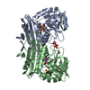

 PDBj
PDBj
 Assembly
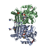

 Mass: 62.068 Da / Num. of mol.: 4 / Source method: obtained synthetically / Formula: C2H6O2
 Mass: 94.971 Da / Num. of mol.: 1 / Source method: obtained synthetically / Formula: PO4
 Mass: 18.015 Da / Num. of mol.: 532 / Source method: isolated from a natural source / Formula: H2O
 Sample preparation
 / Beamline: BL9-2 / Wavelength: 0.91837,0.97951
 Processing