 Movie
Movie Controller


+ Open data

- Basic information
| Entry | Database: PDB / ID: 3lu0 | ||||||
|---|---|---|---|---|---|---|---|
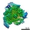
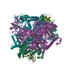
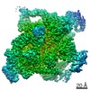
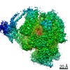
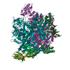
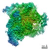
| Title | Molecular model of Escherichia coli core RNA polymerase | ||||||
 Components Components | (DNA-directed RNA polymerase subunit ... Polymerase) x 4 Polymerase) x 4 | ||||||
 Keywords Keywords | TRANSFERASE / E. coli RNA polymerase / DNA-directed RNA polymerase / Nucleotidyltransferase / Transcription | ||||||
| Function / homology |  Function and homology informationRNA polymerase complex / submerged biofilm formation / cellular response to cell envelope stress / cytosolic DNA-directed RNA polymerase complex / regulation of DNA-templated transcription initiation / bacterial-type flagellum assembly / bacterial-type flagellum-dependent cell motility / nitrate assimilation / transcription elongation factor complex / regulation of DNA-templated transcription elongation ...RNA polymerase complex / submerged biofilm formation / cellular response to cell envelope stress / cytosolic DNA-directed RNA polymerase complex / regulation of DNA-templated transcription initiation / bacterial-type flagellum assembly / bacterial-type flagellum-dependent cell motility / nitrate assimilation / transcription elongation factor complex / regulation of DNA-templated transcription elongation / transcription antitermination / cell motility / DNA-templated transcription initiation / ribonucleoside binding / DNA-directed 5'-3' RNA polymerase activity / DNA-directed RNA polymerase / response to heat / protein-containing complex assembly / intracellular iron ion homeostasis / protein dimerization activity / response to antibiotic / magnesium ion binding / DNA binding / zinc ion binding / membrane / cytosol / cytoplasm Function and homology informationRNA polymerase complex / submerged biofilm formation / cellular response to cell envelope stress / cytosolic DNA-directed RNA polymerase complex / regulation of DNA-templated transcription initiation / bacterial-type flagellum assembly / bacterial-type flagellum-dependent cell motility / nitrate assimilation / transcription elongation factor complex / regulation of DNA-templated transcription elongation ...RNA polymerase complex / submerged biofilm formation / cellular response to cell envelope stress / cytosolic DNA-directed RNA polymerase complex / regulation of DNA-templated transcription initiation / bacterial-type flagellum assembly / bacterial-type flagellum-dependent cell motility / nitrate assimilation / transcription elongation factor complex / regulation of DNA-templated transcription elongation / transcription antitermination / cell motility / DNA-templated transcription initiation / ribonucleoside binding / DNA-directed 5'-3' RNA polymerase activity / DNA-directed RNA polymerase / response to heat / protein-containing complex assembly / intracellular iron ion homeostasis / protein dimerization activity / response to antibiotic / magnesium ion binding / DNA binding / zinc ion binding / membrane / cytosol / cytoplasmSimilarity search - Function | ||||||
| Biological species |  Escherichia coli (E. coli) Escherichia coli (E. coli) | ||||||
| Method | ELECTRON MICROSCOPY / single particle reconstruction / cryo EM / Resolution: 11.2 Å | ||||||
 Authors Authors | Darst, S.A. | ||||||
 Citation Citation | Journal: PLoS Biol / Year: 2010 Title: Complete structural model of Escherichia coli RNA polymerase from a hybrid approach. Authors: Natacha Opalka / Jesse Brown / William J Lane / Kelly-Anne F Twist / Robert Landick / Francisco J Asturias / Seth A Darst /  Abstract: The Escherichia coli transcription system is the best characterized from a biochemical and genetic point of view and has served as a model system. Nevertheless, a molecular understanding of the ...The Escherichia coli transcription system is the best characterized from a biochemical and genetic point of view and has served as a model system. Nevertheless, a molecular understanding of the details of E. coli transcription and its regulation, and therefore its full exploitation as a model system, has been hampered by the absence of high-resolution structural information on E. coli RNA polymerase (RNAP). We use a combination of approaches, including high-resolution X-ray crystallography, ab initio structural prediction, homology modeling, and single-particle cryo-electron microscopy, to generate complete atomic models of E. coli core RNAP and an E. coli RNAP ternary elongation complex. The detailed and comprehensive structural descriptions can be used to help interpret previous biochemical and genetic data in a new light and provide a structural framework for designing experiments to understand the function of the E. coli lineage-specific insertions and their role in the E. coli transcription program. | ||||||
| History |
|
- Structure visualization
| Movie |
Movie viewer |
|---|---|
| Structure viewer | Molecule: MolmilJmol/JSmol |
- Downloads & links
-Download
| PDBx/mmCIF format | 3lu0.cif.gz | 565.9 KB | Display | PDBx/mmCIF format |
|---|---|---|---|---|
| PDB format | pdb3lu0.ent.gz | 436.5 KB | Display | PDB format |
| PDBx/mmJSON format | 3lu0.json.gz | Tree view | PDBx/mmJSON format | |
| Others |  Other downloads Other downloads |
-Validation report
| Arichive directory | https://data.pdbj.org/pub/pdb/validation_reports/lu/3lu0ftp://data.pdbj.org/pub/pdb/validation_reports/lu/3lu0 | HTTPS FTP |
|---|
-Related structure data
| Related structure data | 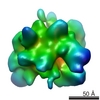 5169MC 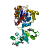 3ltiC M: map data used to model this data C: citing same article ( |
|---|---|
| Similar structure data |


-Links
 PDBj
PDBj

- Assembly
| Deposited unit | 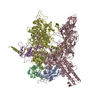
|
|---|---|
| 1 |
|
-Components
-DNA-directed RNA polymerase subunit ... , 4 types, 5 molecules ABCDE
| #1: Protein | Polymerase / RNAP subunit alpha / Transcriptase subunit alpha / RNA polymerase subunit alpha Mass: 36558.680 Da / Num. of mol.: 2 Source method: isolated from a genetically manipulated source Source: (gene. exp.) Escherichia coli (E. coli) / Strain: K12 / Gene: b3295, JW3257, pez, phs, rpoA, sez / Production host: Escherichia coli (E. coli) / References: UniProt: P0A7Z4, DNA-directed RNA polymerase#2: Protein | | Polymerase / RNAP subunit beta / Transcriptase subunit beta / RNA polymerase subunit betaMass: 150804.922 Da / Num. of mol.: 1 Source method: isolated from a genetically manipulated source Source: (gene. exp.) Escherichia coli (E. coli) / Strain: K12Gene: b3987, groN, JW3950, nitB, rif, ron, rpoB, rpoC, stl, stv, tabD Production host: Escherichia coli (E. coli) / References: UniProt: P0A8V2, DNA-directed RNA polymerase#3: Protein | | Polymerase / RNAP subunit beta' / Transcriptase subunit beta' / RNA polymerase subunit beta'Mass: 155366.781 Da / Num. of mol.: 1 Source method: isolated from a genetically manipulated source Source: (gene. exp.) Escherichia coli (E. coli) / Strain: K12 / Gene: b3988, JW3951, rpoC, rpoD, tabB / Production host: Escherichia coli (E. coli) / References: UniProt: P0A8T7, DNA-directed RNA polymerase#4: Protein | | Polymerase / RNAP omega subunit / Transcriptase subunit omega / RNA polymerase omega subunitMass: 10249.547 Da / Num. of mol.: 1 Source method: isolated from a genetically manipulated source Source: (gene. exp.) Escherichia coli (E. coli) / Strain: K12 / Gene: b3649, JW3624, rpoZ / Production host: Escherichia coli (E. coli) / References: UniProt: P0A800, DNA-directed RNA polymerase |
|---|
-Non-polymers , 2 types, 3 molecules 

| #5: Chemical | ChemComp-MG /  Mass: 24.305 Da / Num. of mol.: 1 / Source method: obtained synthetically / Formula: Mg Mass: 24.305 Da / Num. of mol.: 1 / Source method: obtained synthetically / Formula: Mg |
|---|---|
| #6: Chemical |  Mass: 65.409 Da / Num. of mol.: 2 / Source method: obtained synthetically / Formula: Zn Mass: 65.409 Da / Num. of mol.: 2 / Source method: obtained synthetically / Formula: Zn |
-Experimental details
-Experiment
| Experiment | Method: ELECTRON MICROSCOPY |
|---|---|
| EM experiment | Aggregation state: PARTICLE / 3D reconstruction method: single particle reconstruction |
- Sample preparation
| Component | Name: E. coli RNA polymerase / Type: COMPLEX |
|---|---|
| Buffer solution | Name: Tris-HClTris / pH: 8 / Details: Tris-HCl |
| Specimen | Embedding applied: NO / Shadowing applied: NO / Staining applied: NO / Vitrification applied: YES |
| Vitrification | Instrument: HOMEMADE PLUNGER / Cryogen name: ETHANE |
- Electron microscopy imaging
| Experimental equipment |  Model: Tecnai F20 / Image courtesy: FEI Company |
|---|---|
| Microscopy | Model: FEI TECNAI F20 |
| Electron gun | Electron source: FIELD EMISSION GUN / Accelerating voltage: 200 kV / Illumination mode: FLOOD BEAM |
| Electron lens | Mode: BRIGHT FIELDBright-field microscopy / Nominal magnification: 50000 X / Nominal defocus max: 3000 nm / Nominal defocus min: 1800 nm |
| Specimen holder | Specimen holder model: GATAN LIQUID NITROGEN / Specimen holder type: single-tilt / Temperature: 80 K |
| Image recording | Electron dose: 10 e/Å2 / Film or detector model: KODAK SO-163 FILM |
| Image scans | Num. digital images: 48 |
| Radiation | Protocol: SINGLE WAVELENGTH / Monochromatic (M) / Laue (L): M / Scattering type: x-ray |
| Radiation wavelength | Relative weight: 1 |
- Processing
| EM software |
| |||||||||||||||
|---|---|---|---|---|---|---|---|---|---|---|---|---|---|---|---|---|
| Symmetry | Point symmetry: C1 (asymmetric) | |||||||||||||||
| 3D reconstruction | Resolution: 11.2 Å / Num. of particles: 42000 / Nominal pixel size: 2.8 Å / Symmetry type: POINT | |||||||||||||||
| Atomic model building | Protocol: FLEXIBLE FIT / Space: REAL / Details: METHOD--local refinement, flexible fitting | |||||||||||||||
| Refinement step | Cycle: LAST
|
