 Movie
Movie Controller

[English] 日本語
 Yorodumi
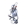
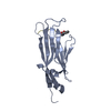
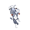
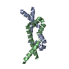
- PDB-3lta: Crystal structure of a non-biological ATP binding protein with a ... -
+ Open data

- Basic information
| Entry | Database: PDB / ID: 3lta | ||||||
|---|---|---|---|---|---|---|---|
| Title | Crystal structure of a non-biological ATP binding protein with a TYR-PHE mutation within the ligand binding domain | ||||||
 Components Components | ATP BINDING PROTEIN-DX | ||||||
 Keywords Keywords |  DE NOVO PROTEIN / ALPHA/BETA FOLD / BENT ATP / NON-BIOLOGICAL PROTEIN DE NOVO PROTEIN / ALPHA/BETA FOLD / BENT ATP / NON-BIOLOGICAL PROTEIN | ||||||
| Function / homology | Nuclear Transport Factor 2; Chain: A, - #210 / Nuclear Transport Factor 2; Chain: A, / Roll / Alpha Beta / ADENOSINE-5'-TRIPHOSPHATE / DI(HYDROXYETHYL)ETHER Function and homology information Function and homology information | ||||||
| Biological species | synthetic construct (others) | ||||||
| Method | X-RAY DIFFRACTION / SYNCHROTRON / MOLECULAR REPLACEMENT / Resolution: 2.7 Å | ||||||
 Authors Authors | Simmons, C.R. / Magee, C.L. / Allen, J.P. / Chaput, J.C. | ||||||
 Citation Citation | Journal: Biochemistry / Year: 2010 Title: Three-dimensional structures reveal multiple ADP/ATP binding modes for a synthetic class of artificial proteins. Authors: Simmons, C.R. / Magee, C.L. / Smith, D.A. / Lauman, L. / Chaput, J.C. / Allen, J.P. | ||||||
| History |
|
- Structure visualization
| Structure viewer | Molecule: MolmilJmol/JSmol |
|---|
- Downloads & links
-Download
| PDBx/mmCIF format | 3lta.cif.gz | 30.6 KB | Display | PDBx/mmCIF format |
|---|---|---|---|---|
| PDB format | pdb3lta.ent.gz | 19.2 KB | Display | PDB format |
| PDBx/mmJSON format | 3lta.json.gz | Tree view | PDBx/mmJSON format | |
| Others |  Other downloads Other downloads |
-Validation report
| Arichive directory | https://data.pdbj.org/pub/pdb/validation_reports/lt/3ltaftp://data.pdbj.org/pub/pdb/validation_reports/lt/3lta | HTTPS FTP |
|---|
-Related structure data
| Related structure data |  3lt8C 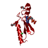 3lt9C 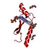 3ltbC 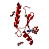 3ltcC 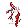 3ltdC  3g4b C: citing same article ( S: Starting model for refinement |
|---|---|
| Similar structure data |


-Links
 PDBj
PDBj
- Assembly
| Deposited unit | 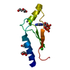
| ||||||||
|---|---|---|---|---|---|---|---|---|---|
| 1 | 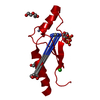
| ||||||||
| Unit cell |
|
-Components
-Protein , 1 types, 1 molecules A
| #1: Protein | Mass: 9692.112 Da / Num. of mol.: 1 Source method: isolated from a genetically manipulated source Source: (gene. exp.) synthetic construct (others) Description: THE PROTEIN IS A NON-BIOLOGICAL PROTEIN SELECTED FROM A RANDOM POOL OF PROTEIN SEQUENCES FOR THE ABILITY TO BIND ATP Plasmid: PIADL14 / Production host:  Escherichia coli (E. coli) / Strain (production host): BL21(DE3) Escherichia coli (E. coli) / Strain (production host): BL21(DE3) |
|---|
-Non-polymers , 5 types, 25 molecules 


| #2: Chemical | ChemComp-ZN /  Mass: 65.409 Da / Num. of mol.: 1 / Source method: obtained synthetically / Formula: Zn Mass: 65.409 Da / Num. of mol.: 1 / Source method: obtained synthetically / Formula: Zn | ||||
|---|---|---|---|---|---|
| #3: Chemical | ChemComp-ATP / Adenosine triphosphate Mass: 507.181 Da / Num. of mol.: 1 / Source method: obtained synthetically / Formula: C10H16N5O13P3 / Comment: ATP, energy-carrying molecule*YM Mass: 507.181 Da / Num. of mol.: 1 / Source method: obtained synthetically / Formula: C10H16N5O13P3 / Comment: ATP, energy-carrying molecule*YM | ||||
| #4: Chemical | Diethylene glycol Mass: 106.120 Da / Num. of mol.: 3 / Source method: obtained synthetically / Formula: C4H10O3 Mass: 106.120 Da / Num. of mol.: 3 / Source method: obtained synthetically / Formula: C4H10O3#5: Chemical | ChemComp-CL / | Chloride Mass: 35.453 Da / Num. of mol.: 1 / Source method: obtained synthetically / Formula: Cl Mass: 35.453 Da / Num. of mol.: 1 / Source method: obtained synthetically / Formula: Cl#6: Water | ChemComp-HOH / | WaterMass: 18.015 Da / Num. of mol.: 19 / Source method: isolated from a natural source / Formula: H2O |
-Experimental details
-Experiment
| Experiment | Method: X-RAY DIFFRACTION / Number of used crystals: 1 |
|---|
- Sample preparation
| Crystal | Density Matthews: 4.21 Å3/Da / Density % sol: 70.8 % |
|---|---|
| Crystal grow | pH: 8.5 Details: 0.1 M SODIUM PHOSPHATE PH 8.5, 0.25 M SODIUM CITRATE, 0.3 M SODIUM CHLORIDE, 23% PEG 400, VAPOR DIFFUSION, SITTING DROP, TEMPERATURE 298K |
-Data collection
| Diffraction | Mean temperature: 100 K |
|---|---|
| Diffraction source | Source: SYNCHROTRON / Site: NSLS  / Beamline: X12B / Wavelength: 1.2817 / Beamline: X12B / Wavelength: 1.2817 |
| Detector | Type: ADSC QUANTUM 4 / Detector: CCD / Date: Jun 18, 2008 |
| Radiation | Monochromator: SI(111) / Protocol: SINGLE WAVELENGTH / Monochromatic (M) / Laue (L): M / Scattering type: x-ray |
| Radiation wavelength | Wavelength: 1.2817 Å / Relative weight: 1 |
| Reflection | Resolution: 2.7→50 Å / Num. obs: 4710 / % possible obs: 99.3 % / Redundancy: 4.2 % / Rmerge(I) obs: 0.135 / Net I/σ(I): 15.385 |
| Reflection shell | Resolution: 2.7→2.8 Å / Redundancy: 4.2 % / Rmerge(I) obs: 0.57 / % possible all: 99.4 |
- Processing
| Software |
| ||||||||||||||||||||||||||||||||||||||||||||||||||||||||||||||||||||||||||||||||||||||||||||||||||||||||||||||||||||||||||||||||||||||||||||||||||||||||||||||||||||||||||
|---|---|---|---|---|---|---|---|---|---|---|---|---|---|---|---|---|---|---|---|---|---|---|---|---|---|---|---|---|---|---|---|---|---|---|---|---|---|---|---|---|---|---|---|---|---|---|---|---|---|---|---|---|---|---|---|---|---|---|---|---|---|---|---|---|---|---|---|---|---|---|---|---|---|---|---|---|---|---|---|---|---|---|---|---|---|---|---|---|---|---|---|---|---|---|---|---|---|---|---|---|---|---|---|---|---|---|---|---|---|---|---|---|---|---|---|---|---|---|---|---|---|---|---|---|---|---|---|---|---|---|---|---|---|---|---|---|---|---|---|---|---|---|---|---|---|---|---|---|---|---|---|---|---|---|---|---|---|---|---|---|---|---|---|---|---|---|---|---|---|---|---|
| Refinement | Method to determine structure: MOLECULAR REPLACEMENT Starting model: PDB ENTRY 3G4B 3g4b Resolution: 2.7→41.42 Å / Cor.coef. Fo:Fc: 0.925 / Cor.coef. Fo:Fc free: 0.905 / SU B: 10.212 / SU ML: 0.197 / Cross valid method: THROUGHOUT / σ(F): 0 / ESU R: 0.328 / ESU R Free: 0.25 / Stereochemistry target values: MAXIMUM LIKELIHOOD Details: HYDROGENS HAVE BEEN ADDED IN THE RIDING POSITIONS. U VALUES: REFINED INDIVIDUALLY
| ||||||||||||||||||||||||||||||||||||||||||||||||||||||||||||||||||||||||||||||||||||||||||||||||||||||||||||||||||||||||||||||||||||||||||||||||||||||||||||||||||||||||||
| Solvent computation | Ion probe radii: 0.8 Å / Shrinkage radii: 0.8 Å / VDW probe radii: 1.2 Å / Solvent model: MASK | ||||||||||||||||||||||||||||||||||||||||||||||||||||||||||||||||||||||||||||||||||||||||||||||||||||||||||||||||||||||||||||||||||||||||||||||||||||||||||||||||||||||||||
| Displacement parameters | Biso mean: 32.69 Å2
| ||||||||||||||||||||||||||||||||||||||||||||||||||||||||||||||||||||||||||||||||||||||||||||||||||||||||||||||||||||||||||||||||||||||||||||||||||||||||||||||||||||||||||
| Refinement step | Cycle: LAST / Resolution: 2.7→41.42 Å
| ||||||||||||||||||||||||||||||||||||||||||||||||||||||||||||||||||||||||||||||||||||||||||||||||||||||||||||||||||||||||||||||||||||||||||||||||||||||||||||||||||||||||||
| Refine LS restraints |
| ||||||||||||||||||||||||||||||||||||||||||||||||||||||||||||||||||||||||||||||||||||||||||||||||||||||||||||||||||||||||||||||||||||||||||||||||||||||||||||||||||||||||||
| LS refinement shell | Resolution: 2.7→2.77 Å / Total num. of bins used: 20
|
