 Movie
Movie Controller


+ Open data

- Basic information
| Entry | Database: PDB / ID: 3ljp | ||||||
|---|---|---|---|---|---|---|---|
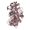
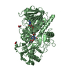
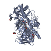
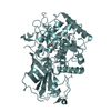
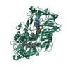
| Title | Crystal structure of choline oxidase V464A mutant | ||||||
 Components Components | Choline oxidase | ||||||
 Keywords Keywords | OXIDOREDUCTASE / Flavoenzyme Oxidase / Covalently linked FAD / FAD / Flavoprotein | ||||||
| Function / homology |  Function and homology informationcholine oxidase / choline:oxygen 1-oxidoreductase activity / glycine betaine biosynthetic process from choline / flavin adenine dinucleotide binding Function and homology informationcholine oxidase / choline:oxygen 1-oxidoreductase activity / glycine betaine biosynthetic process from choline / flavin adenine dinucleotide bindingSimilarity search - Function | ||||||
| Biological species |  Arthrobacter globiformis (bacteria) Arthrobacter globiformis (bacteria) | ||||||
| Method | X-RAY DIFFRACTION / SYNCHROTRON / MOLECULAR REPLACEMENT / Resolution: 2.2 Å | ||||||
 Authors Authors | Finnegan, S. / Agniswamy, J. / Weber, I.T. / Giovanni, G. | ||||||
 Citation Citation | Journal: Biochemistry / Year: 2010 Title: Role of valine 464 in the flavin oxidation reaction catalyzed by choline oxidase. Authors: Finnegan, S. / Agniswamy, J. / Weber, I.T. / Gadda, G. | ||||||
| History |
|
- Structure visualization
| Structure viewer | Molecule: MolmilJmol/JSmol |
|---|
- Downloads & links
-Download
| PDBx/mmCIF format | 3ljp.cif.gz | 225.5 KB | Display | PDBx/mmCIF format |
|---|---|---|---|---|
| PDB format | pdb3ljp.ent.gz | 179.9 KB | Display | PDB format |
| PDBx/mmJSON format | 3ljp.json.gz | Tree view | PDBx/mmJSON format | |
| Others |  Other downloads Other downloads |
-Validation report
| Arichive directory | https://data.pdbj.org/pub/pdb/validation_reports/lj/3ljpftp://data.pdbj.org/pub/pdb/validation_reports/lj/3ljp | HTTPS FTP |
|---|
-Related structure data
| Related structure data |  2jbvS S: Starting model for refinement |
|---|---|
| Similar structure data |


-Links
 PDBj
PDBj
- Assembly
| Deposited unit | 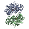
| ||||||||
|---|---|---|---|---|---|---|---|---|---|
| 1 | 
| ||||||||
| 2 | 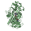
| ||||||||
| Unit cell |
|
-Components
| #1: Protein | Mass: 59870.707 Da / Num. of mol.: 2 / Mutation: V464A Source method: isolated from a genetically manipulated source Source: (gene. exp.) Arthrobacter globiformis (bacteria) / Gene: codA / Production host: Escherichia coli (E. coli) / Strain (production host): JM109 / References: UniProt: Q7X2H8, choline oxidase#2: Chemical |   Mass: 787.566 Da / Num. of mol.: 2 / Source method: obtained synthetically / Formula: C27H35N9O15P2 Mass: 787.566 Da / Num. of mol.: 2 / Source method: obtained synthetically / Formula: C27H35N9O15P2#3: Water | ChemComp-HOH / | Water Mass: 18.015 Da / Num. of mol.: 472 / Source method: isolated from a natural source / Formula: H2O Mass: 18.015 Da / Num. of mol.: 472 / Source method: isolated from a natural source / Formula: H2O |
|---|
-Experimental details
-Experiment
| Experiment | Method: X-RAY DIFFRACTION / Number of used crystals: 1 |
|---|
- Sample preparation
| Crystal | Density Matthews: 2.79 Å3/Da / Density % sol: 55.95 % |
|---|---|
| Crystal grow | Temperature: 298 K / Method: vapor diffusion, hanging drop / pH: 6 Details: 10-15% PEG 6000, 50-200mM magnesium acetate, 200mM trimethylamine, 0.08M sodium cacodilate, pH 6.0, VAPOR DIFFUSION, HANGING DROP, temperature 298K |
-Data collection
| Diffraction | Mean temperature: 100 K |
|---|---|
| Diffraction source | Source: SYNCHROTRON / Site: APS  / Beamline: 22-ID / Wavelength: 0.8 Å / Beamline: 22-ID / Wavelength: 0.8 Å |
| Detector | Type: MAR scanner 300 mm plate / Detector: IMAGE PLATE / Date: Jun 10, 2009 / Details: mirrors |
| Radiation | Protocol: SINGLE WAVELENGTH / Monochromatic (M) / Laue (L): M / Scattering type: x-ray |
| Radiation wavelength | Wavelength: 0.8 Å / Relative weight: 1 |
| Reflection | Resolution: 2.2→50 Å / Num. all: 70579 / Num. obs: 67756 / % possible obs: 96 % / Observed criterion σ(F): 0 / Observed criterion σ(I): 2 / Redundancy: 6.1 % / Rmerge(I) obs: 0.134 / Net I/σ(I): 12.31 |
| Reflection shell | Resolution: 2.2→2.28 Å / Redundancy: 6.6 % / Rmerge(I) obs: 0.838 / Mean I/σ(I) obs: 5.51 / Num. unique all: 6854 / % possible all: 99.5 |
- Processing
| Software |
| |||||||||||||||||||||||||||||||||||||||||||||||||||||||||||||||||||||||||||
|---|---|---|---|---|---|---|---|---|---|---|---|---|---|---|---|---|---|---|---|---|---|---|---|---|---|---|---|---|---|---|---|---|---|---|---|---|---|---|---|---|---|---|---|---|---|---|---|---|---|---|---|---|---|---|---|---|---|---|---|---|---|---|---|---|---|---|---|---|---|---|---|---|---|---|---|---|
| Refinement | Method to determine structure: MOLECULAR REPLACEMENT Starting model: PDB ENTRY 2JBV Resolution: 2.2→43.52 Å / Cor.coef. Fo:Fc: 0.949 / Cor.coef. Fo:Fc free: 0.913 / SU B: 9.196 / SU ML: 0.109 / TLS residual ADP flag: LIKELY RESIDUAL / Cross valid method: THROUGHOUT / σ(F): 0 / ESU R: 0.222 / ESU R Free: 0.197 / Stereochemistry target values: MAXIMUM LIKELIHOOD / Details: HYDROGENS HAVE BEEN ADDED IN THE RIDING POSITIONS
| |||||||||||||||||||||||||||||||||||||||||||||||||||||||||||||||||||||||||||
| Solvent computation | Ion probe radii: 0.8 Å / Shrinkage radii: 0.8 Å / VDW probe radii: 1.4 Å / Solvent model: MASK | |||||||||||||||||||||||||||||||||||||||||||||||||||||||||||||||||||||||||||
| Displacement parameters | Biso mean: 19.429 Å2
| |||||||||||||||||||||||||||||||||||||||||||||||||||||||||||||||||||||||||||
| Refinement step | Cycle: LAST / Resolution: 2.2→43.52 Å
| |||||||||||||||||||||||||||||||||||||||||||||||||||||||||||||||||||||||||||
| Refine LS restraints |
| |||||||||||||||||||||||||||||||||||||||||||||||||||||||||||||||||||||||||||
| LS refinement shell | Resolution: 2.196→2.252 Å / Total num. of bins used: 20
| |||||||||||||||||||||||||||||||||||||||||||||||||||||||||||||||||||||||||||
| Refinement TLS params. | Method: refined / Refine-ID: X-RAY DIFFRACTION
| |||||||||||||||||||||||||||||||||||||||||||||||||||||||||||||||||||||||||||
| Refinement TLS group |
|
