 Movie
Movie Controller


+ Open data

- Basic information
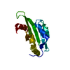
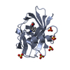
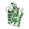
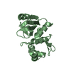
| Entry | Database: PDB / ID: 3lj8 | ||||||
|---|---|---|---|---|---|---|---|
| Title | Crystal Structure of MKP-4 | ||||||
 Components Components | Tyrosine-protein phosphatase | ||||||
 Keywords Keywords |  HYDROLASE / alpha/beta hydrolase / Protein phosphatase HYDROLASE / alpha/beta hydrolase / Protein phosphatase | ||||||
| Function / homology |  Function and homology information Function and homology informationMAP kinase tyrosine phosphatase activity / protein tyrosine/threonine phosphatase activity / MAP kinase tyrosine/serine/threonine phosphatase activity / protein tyrosine/serine/threonine phosphatase activity / Signaling by MAPK mutants / RAF-independent MAPK1/3 activation / negative regulation of MAPK cascade / myosin phosphatase activity / protein-serine/threonine phosphatase / phosphoprotein phosphatase activity ...MAP kinase tyrosine phosphatase activity / protein tyrosine/threonine phosphatase activity / MAP kinase tyrosine/serine/threonine phosphatase activity / protein tyrosine/serine/threonine phosphatase activity / Signaling by MAPK mutants / RAF-independent MAPK1/3 activation / negative regulation of MAPK cascade / myosin phosphatase activity / protein-serine/threonine phosphatase / phosphoprotein phosphatase activity / JNK cascade / ERK1 and ERK2 cascade / protein dephosphorylation / protein-tyrosine-phosphatase / Negative regulation of MAPK pathway / MAPK cascade / nucleus / cytosol / cytoplasmSimilarity search - Function | ||||||
| Biological species |  Homo sapiens (human) Homo sapiens (human) | ||||||
| Method | X-RAY DIFFRACTION / SYNCHROTRON / MOLECULAR REPLACEMENT / Resolution: 2.7 Å | ||||||
 Authors Authors | Jeong, D.G. / Yoon, T.S. / Jung, S.-K. / Park, H.S. / Ryu, S.E. / Kim, S.J. | ||||||
 Citation Citation | Journal: Acta Crystallogr.,Sect.D / Year: 2011 Title: Exploring binding sites other than the catalytic core in the crystal structure of the catalytic domain of MKP-4 Authors: Jeong, D.G. / Yoon, T.S. / Jung, S.-K. / Park, B.C. / Park, H.S. / Ryu, S.E. / Kim, S.J. | ||||||
| History |
|
- Structure visualization
| Structure viewer | Molecule: MolmilJmol/JSmol |
|---|
- Downloads & links
-Download
| PDBx/mmCIF format | 3lj8.cif.gz | 40.8 KB | Display | PDBx/mmCIF format |
|---|---|---|---|---|
| PDB format | pdb3lj8.ent.gz | 28.4 KB | Display | PDB format |
| PDBx/mmJSON format | 3lj8.json.gz | Tree view | PDBx/mmJSON format | |
| Others |  Other downloads Other downloads |
-Validation report
| Arichive directory | https://data.pdbj.org/pub/pdb/validation_reports/lj/3lj8ftp://data.pdbj.org/pub/pdb/validation_reports/lj/3lj8 | HTTPS FTP |
|---|
-Related structure data
| Related structure data |  1zzwS S: Starting model for refinement |
|---|---|
| Similar structure data |


-Links
 PDBj
PDBj


- Assembly
| Deposited unit | 
| ||||||||
|---|---|---|---|---|---|---|---|---|---|
| 1 |
| ||||||||
| Unit cell |
|
-Components
| #1: Protein | Mass: 16652.029 Da / Num. of mol.: 1 / Fragment: catalytic domain, residues 202-347 Source method: isolated from a genetically manipulated source Source: (gene. exp.) Homo sapiens (human) / Plasmid: pET28a / Production host:  Escherichia coli (E. coli) Escherichia coli (E. coli)References: UniProt: Q99956, protein-tyrosine-phosphatase, protein-serine/threonine phosphatase |
|---|
-Experimental details
-Experiment
| Experiment | Method: X-RAY DIFFRACTION / Number of used crystals: 1 |
|---|
- Sample preparation
| Crystal | Density Matthews: 4.15 Å3/Da / Density % sol: 70.33 % |
|---|---|
| Crystal grow | Temperature: 291 K / Method: vapor diffusion, hanging drop / pH: 7.5 Details: 10% ethanol, 1.3M NaCl, 0.2M MgSO4, pH 7.5, VAPOR DIFFUSION, HANGING DROP, temperature 291K |
-Data collection
| Diffraction | Mean temperature: 93 K |
|---|---|
| Diffraction source | Source: SYNCHROTRON / Site: Photon Factory  / Beamline: BL-5A / Wavelength: 1 Å / Beamline: BL-5A / Wavelength: 1 Å |
| Detector | Type: ADSC QUANTUM 315 / Detector: CCD / Date: May 21, 2005 / Details: mirrors |
| Radiation | Monochromator: mirrors / Protocol: SINGLE WAVELENGTH / Monochromatic (M) / Laue (L): M / Scattering type: x-ray |
| Radiation wavelength | Wavelength: 1 Å / Relative weight: 1 |
| Reflection | Resolution: 2.7→50 Å / Num. all: 7864 / Num. obs: 7730 / % possible obs: 98.3 % / Observed criterion σ(F): 0 / Observed criterion σ(I): 0 / Redundancy: 5.7 % / Rmerge(I) obs: 0.032 / Net I/σ(I): 14.7 |
| Reflection shell | Resolution: 2.7→2.85 Å / Redundancy: 6 % / Rmerge(I) obs: 0.228 / Mean I/σ(I) obs: 3.4 / % possible all: 98.5 |
- Processing
| Software |
| |||||||||||||||||||||||||
|---|---|---|---|---|---|---|---|---|---|---|---|---|---|---|---|---|---|---|---|---|---|---|---|---|---|---|
| Refinement | Method to determine structure: MOLECULAR REPLACEMENT Starting model: PDB entry 1zzw Resolution: 2.7→35.68 Å / Isotropic thermal model: isotropic / Cross valid method: THROUGHOUT / σ(F): 0 / σ(I): 0 / Stereochemistry target values: Engh & Huber
| |||||||||||||||||||||||||
| Displacement parameters | Biso mean: 67.3 Å2 | |||||||||||||||||||||||||
| Refinement step | Cycle: LAST / Resolution: 2.7→35.68 Å
| |||||||||||||||||||||||||
| Refine LS restraints |
|
