 Movie
Movie Controller

[English] 日本語
 Yorodumi
- PDB-3l9i: Myosin VI nucleotide-free (mdinsert2) L310G mutant crystal structure -
+ Open data

- Basic information
| Entry | Database: PDB / ID: 3l9i | ||||||
|---|---|---|---|---|---|---|---|
| Title | Myosin VI nucleotide-free (mdinsert2) L310G mutant crystal structure | ||||||
 Components Components |
| ||||||
 Keywords Keywords |  MOTOR PROTEIN / myosin VI / unconventional myosin / directionality / motility / gating / Actin-binding / ATP-binding / Calmodulin-binding / Endocytosis / Golgi apparatus / Hearing / Membrane / Myosin / Nucleotide-binding / Nucleus / Phosphoprotein / Protein transport / Transport MOTOR PROTEIN / myosin VI / unconventional myosin / directionality / motility / gating / Actin-binding / ATP-binding / Calmodulin-binding / Endocytosis / Golgi apparatus / Hearing / Membrane / Myosin / Nucleotide-binding / Nucleus / Phosphoprotein / Protein transport / Transport | ||||||
| Function / homology |  Function and homology information Function and homology informationnegative regulation of phospholipase C-activating phototransduction signaling pathway / myosin VI complex / myosin VI head/neck binding / myosin VII complex / photoreceptor cell axon guidance / negative regulation of opsin-mediated signaling pathway / rhabdomere / rhabdomere development / myosin V complex / : ...negative regulation of phospholipase C-activating phototransduction signaling pathway / myosin VI complex / myosin VI head/neck binding / myosin VII complex / photoreceptor cell axon guidance / negative regulation of opsin-mediated signaling pathway / rhabdomere / rhabdomere development / myosin V complex / : / regulation of secretion / kinetochore organization / : / actin filament-based movement / G protein-coupled opsin signaling pathway / Neutrophil degranulation / inner ear auditory receptor cell differentiation / myosin V binding / channel regulator activity / vesicle transport along actin filament / cellular response to ethanol / myosin complex / clathrin-coated vesicle / microfilament motor activity / inner ear morphogenesis / muscle cell cellular homeostasis / myosin heavy chain binding / mitotic spindle pole / filamentous actin / microvillus / centriole replication / cytoskeletal motor activity / DNA damage response, signal transduction by p53 class mediator / enzyme regulator activity / ruffle / centriole / filopodium / actin filament organization / actin filament / ADP binding / sensory perception of sound / intracellular protein transport / mitotic spindle / spindle / ruffle membrane / endocytosis / actin filament binding / sensory perception of smell / actin cytoskeleton / cell cortex / midbody / cytoplasmic vesicle / nuclear membrane / vesicle / calmodulin binding / protein phosphorylation / centrosome / calcium ion binding / perinuclear region of cytoplasm / Golgi apparatus / nucleoplasm / ATP binding / nucleus / cytosol / cytoplasmSimilarity search - Function | ||||||
| Biological species |  Sus scrofa (pig) Sus scrofa (pig) Drosophila melanogaster (fruit fly) Drosophila melanogaster (fruit fly) | ||||||
| Method | X-RAY DIFFRACTION / SYNCHROTRON / MOLECULAR REPLACEMENT / Resolution: 2.2 Å | ||||||
 Authors Authors | Pylypenko, O. / Song, L. / Sweeney, L.H. / Houdusse, A. | ||||||
 Citation Citation | Journal: To be Published Title: Role of insert i of myosin VI in modulating nucleotide affinity Authors: Pylypenko, O. / Song, L. / Squires, G. / Liu, X. / Zong, A.B. / Houdusse, A. / Sweeney, L.H. | ||||||
| History |
|
- Structure visualization
| Structure viewer | Molecule: MolmilJmol/JSmol |
|---|
- Downloads & links
-Download
| PDBx/mmCIF format | 3l9i.cif.gz | 227.5 KB | Display | PDBx/mmCIF format |
|---|---|---|---|---|
| PDB format | pdb3l9i.ent.gz | 176.9 KB | Display | PDB format |
| PDBx/mmJSON format | 3l9i.json.gz | Tree view | PDBx/mmJSON format | |
| Others |  Other downloads Other downloads |
-Validation report
| Arichive directory | https://data.pdbj.org/pub/pdb/validation_reports/l9/3l9iftp://data.pdbj.org/pub/pdb/validation_reports/l9/3l9i | HTTPS FTP |
|---|
-Related structure data
| Related structure data | 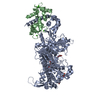 2bkhS S: Starting model for refinement |
|---|---|
| Similar structure data |


-Links
 PDBj
PDBj


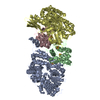
- Assembly
| Deposited unit | 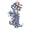
| ||||||||
|---|---|---|---|---|---|---|---|---|---|
| 1 |
| ||||||||
| Unit cell |
|
-Components
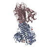
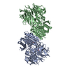
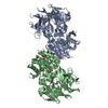
-Protein , 2 types, 2 molecules AC
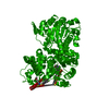
| #1: Protein | / Unconventional myosin VI Mass: 92976.969 Da / Num. of mol.: 1 / Fragment: MOTOR DOMAIN-INSERT2, RESIDUES 2-816 / Mutation: L310G Source method: isolated from a genetically manipulated source Source: (gene. exp.) Sus scrofa (pig) / Gene: MYO6 / Production host:  Spodoptera frugiperda (fall armyworm) / References: UniProt: Q29122 Spodoptera frugiperda (fall armyworm) / References: UniProt: Q29122 |
|---|---|
| #2: Protein | / CaM Mass: 16825.520 Da / Num. of mol.: 1 Source method: isolated from a genetically manipulated source Source: (gene. exp.) Drosophila melanogaster (fruit fly) / Gene: Cam, CG8472 / Production host: Spodoptera frugiperda (fall armyworm) / References: UniProt: P62152 |
-Non-polymers , 5 types, 840 molecules 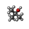


| #3: Chemical | ChemComp-TBU / Tert-Butyl alcohol Mass: 74.122 Da / Num. of mol.: 1 / Source method: obtained synthetically / Formula: C4H10O Mass: 74.122 Da / Num. of mol.: 1 / Source method: obtained synthetically / Formula: C4H10O | ||||||
|---|---|---|---|---|---|---|---|
| #4: Chemical | Acetate Mass: 59.044 Da / Num. of mol.: 2 / Source method: obtained synthetically / Formula: C2H3O2 Mass: 59.044 Da / Num. of mol.: 2 / Source method: obtained synthetically / Formula: C2H3O2#5: Chemical | ChemComp-PO4 / Phosphate Mass: 94.971 Da / Num. of mol.: 4 / Source method: obtained synthetically / Formula: PO4 Mass: 94.971 Da / Num. of mol.: 4 / Source method: obtained synthetically / Formula: PO4#6: Chemical | ChemComp-CA /  Mass: 40.078 Da / Num. of mol.: 4 / Source method: obtained synthetically / Formula: Ca Mass: 40.078 Da / Num. of mol.: 4 / Source method: obtained synthetically / Formula: Ca#7: Water | ChemComp-HOH / | WaterMass: 18.015 Da / Num. of mol.: 829 / Source method: isolated from a natural source / Formula: H2O |
-Details
| Sequence details | THE AUTHORS STATE THAT THE ORIGINAL SEQUENCE (UNIPROT Q29122) OF MYOSIN VI FROM PIG WAS MOST LIKELY ...THE AUTHORS STATE THAT THE ORIGINAL SEQUENCE (UNIPROT Q29122) OF MYOSIN VI FROM PIG WAS MOST LIKELY INCORRECT BECAUSE THE CHANGES THAT ARE IN THEIR CLONE (LYS DELETION AND THE 6 MUTATIONS) ARE CONSERVED ACROSS THE MYOSIN VI FAMILY. |
|---|
-Experimental details
-Experiment
| Experiment | Method: X-RAY DIFFRACTION / Number of used crystals: 1 |
|---|
- Sample preparation
| Crystal | Density Matthews: 2.94 Å3/Da / Density % sol: 58.2 % |
|---|---|
| Crystal grow | Temperature: 277 K / Method: vapor diffusion, hanging drop / pH: 9 Details: 4% PEG 8000, 50mM Glycine pH9, 3% iso-propanol, 3% tert-butanol, 1mM TCEP , VAPOR DIFFUSION, HANGING DROP, temperature 277K |
-Data collection
| Diffraction | Mean temperature: 100 K |
|---|---|
| Diffraction source | Source: SYNCHROTRON / Site: SOLEIL  / Beamline: PROXIMA 1 / Wavelength: 1.1271 Å / Beamline: PROXIMA 1 / Wavelength: 1.1271 Å |
| Detector | Type: ADSC QUANTUM 315r / Detector: CCD / Date: Apr 30, 2008 |
| Radiation | Monochromator: Kirkpatrick-Baez pair of bi-morph mirrors plus channel cut cryogenically cooled monochromator crystal Protocol: SINGLE WAVELENGTH / Monochromatic (M) / Laue (L): M / Scattering type: x-ray |
| Radiation wavelength | Wavelength: 1.1271 Å / Relative weight: 1 |
| Reflection | Resolution: 2.2→19.97 Å / Num. all: 64550 / Num. obs: 64176 / % possible obs: 99.4 % / Observed criterion σ(F): -3 / Observed criterion σ(I): -3 / Redundancy: 3.9 % / Biso Wilson estimate: 41.5 Å2 / Rsym value: 0.061 / Net I/σ(I): 14.25 |
| Reflection shell | Resolution: 2.2→2.25 Å / Redundancy: 3.9 % / Mean I/σ(I) obs: 3.77 / Num. unique all: 4157 / Rsym value: 0.38 / % possible all: 99.9 |
- Processing
| Software |
| |||||||||||||||||||||||||||||||||||||||||||||||||||||||||||||||||||||||||||||||||||||
|---|---|---|---|---|---|---|---|---|---|---|---|---|---|---|---|---|---|---|---|---|---|---|---|---|---|---|---|---|---|---|---|---|---|---|---|---|---|---|---|---|---|---|---|---|---|---|---|---|---|---|---|---|---|---|---|---|---|---|---|---|---|---|---|---|---|---|---|---|---|---|---|---|---|---|---|---|---|---|---|---|---|---|---|---|---|---|
| Refinement | Method to determine structure: MOLECULAR REPLACEMENT Starting model: 2BKH Resolution: 2.2→19.97 Å / Cor.coef. Fo:Fc: 0.952 / Cor.coef. Fo:Fc free: 0.923 / SU B: 5.744 / SU ML: 0.15 / Cross valid method: THROUGHOUT / ESU R: 0.234 / ESU R Free: 0.197 / Stereochemistry target values: MAXIMUM LIKELIHOOD
| |||||||||||||||||||||||||||||||||||||||||||||||||||||||||||||||||||||||||||||||||||||
| Solvent computation | Ion probe radii: 0.8 Å / Shrinkage radii: 0.8 Å / VDW probe radii: 1.2 Å / Solvent model: BABINET MODEL WITH MASK | |||||||||||||||||||||||||||||||||||||||||||||||||||||||||||||||||||||||||||||||||||||
| Displacement parameters | Biso mean: 40.942 Å2 | |||||||||||||||||||||||||||||||||||||||||||||||||||||||||||||||||||||||||||||||||||||
| Refinement step | Cycle: LAST / Resolution: 2.2→19.97 Å
| |||||||||||||||||||||||||||||||||||||||||||||||||||||||||||||||||||||||||||||||||||||
| Refine LS restraints |
| |||||||||||||||||||||||||||||||||||||||||||||||||||||||||||||||||||||||||||||||||||||
| LS refinement shell | Resolution: 2.2→2.256 Å / Total num. of bins used: 20
|
