 Movie
Movie Controller

[English] 日本語
 Yorodumi
- PDB-3l2o: Structure-Based Mechanism of Dimerization-Dependent Ubiquitinatio... -
+ Open data

- Basic information
| Entry | Database: PDB / ID: 3l2o | ||||||
|---|---|---|---|---|---|---|---|
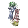
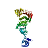
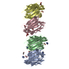
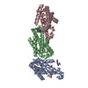
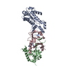
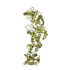
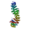
| Title | Structure-Based Mechanism of Dimerization-Dependent Ubiquitination by the SCFFbx4 Ubiquitin Ligase | ||||||
 Components Components |
| ||||||
 Keywords Keywords | PROTEIN BINDING/CELL CYCLE / small G protein fold / Ubl conjugation pathway /  ubiquitin protein ligase / PROTEIN BINDING-CELL CYCLE complex ubiquitin protein ligase / PROTEIN BINDING-CELL CYCLE complex | ||||||
| Function / homology |  Function and homology information Function and homology informationpositive regulation of protein polyubiquitination / F-box domain binding / common myeloid progenitor cell proliferation / PcG protein complex / cellular homeostasis / regulation of DNA damage checkpoint / Cul7-RING ubiquitin ligase complex / positive regulation of ubiquitin protein ligase activity / maintenance of protein location in nucleus / Loss of Function of FBXW7 in Cancer and NOTCH1 Signaling ...positive regulation of protein polyubiquitination / F-box domain binding / common myeloid progenitor cell proliferation / PcG protein complex / cellular homeostasis / regulation of DNA damage checkpoint / Cul7-RING ubiquitin ligase complex / positive regulation of ubiquitin protein ligase activity / maintenance of protein location in nucleus / Loss of Function of FBXW7 in Cancer and NOTCH1 Signaling / negative regulation of protein localization to nucleus / SCF-dependent proteasomal ubiquitin-dependent protein catabolic process / SCF ubiquitin ligase complex / ubiquitin ligase complex scaffold activity / post-transcriptional regulation of gene expression / Prolactin receptor signaling / protein monoubiquitination / Association of TriC/CCT with target proteins during biosynthesis / cullin family protein binding / ubiquitin-like ligase-substrate adaptor activity / protein K48-linked ubiquitination / Nuclear events stimulated by ALK signaling in cancer / ubiquitin ligase complex / negative regulation of fibroblast proliferation / positive regulation of telomere maintenance via telomerase / telomere maintenance / Regulation of BACH1 activity / MAP3K8 (TPL2)-dependent MAPK1/3 activation / positive regulation of protein ubiquitination / SCF-beta-TrCP mediated degradation of Emi1 / NIK-->noncanonical NF-kB signaling / molecular function activator activity / Vpu mediated degradation of CD4 / cellular response to ionizing radiation / Dectin-1 mediated noncanonical NF-kB signaling / Degradation of GLI1 by the proteasome / Activation of NF-kappaB in B cells / Negative regulation of NOTCH4 signaling / Iron uptake and transport / GSK3B and BTRC:CUL1-mediated-degradation of NFE2L2 / Degradation of GLI2 by the proteasome / GLI3 is processed to GLI3R by the proteasome / FBXL7 down-regulates AURKA during mitotic entry and in early mitosis / protein destabilization / regulation of protein stability / Degradation of beta-catenin by the destruction complex / NOTCH1 Intracellular Domain Regulates Transcription / CLEC7A (Dectin-1) signaling / SCF(Skp2)-mediated degradation of p27/p21 / beta-catenin binding / Constitutive Signaling by NOTCH1 PEST Domain Mutants / Constitutive Signaling by NOTCH1 HD+PEST Domain Mutants / FCERI mediated NF-kB activation / Interleukin-1 signaling / protein polyubiquitination / Orc1 removal from chromatin / ubiquitin-protein transferase activity / Regulation of RUNX2 expression and activity / Cyclin D associated events in G1 / ubiquitin protein ligase activity / Regulation of PLK1 Activity at G2/M Transition / cellular senescence / Antigen processing: Ubiquitination & Proteasome degradation / Circadian Clock / Downstream TCR signaling / Neddylation / ubiquitin-dependent protein catabolic process / proteasome-mediated ubiquitin-dependent protein catabolic process / protein ubiquitination / chromatin remodeling / protein domain specific binding / centrosome / protein homodimerization activity / nucleoplasm / nucleus / cytosol / cytoplasmSimilarity search - Function | ||||||
| Biological species |  Homo sapiens (human) Homo sapiens (human) | ||||||
| Method | X-RAY DIFFRACTION / SYNCHROTRON / SAD / Resolution: 2.8 Å | ||||||
 Authors Authors | Li, Y. / Hao, B. | ||||||
 Citation Citation | Journal: J.Biol.Chem. / Year: 2010 Title: Structural basis of dimerization-dependent ubiquitination by the SCF(Fbx4) ubiquitin ligase. Authors: Li, Y. / Hao, B. | ||||||
| History |
|
- Structure visualization
| Structure viewer | Molecule: MolmilJmol/JSmol |
|---|
- Downloads & links
-Download
| PDBx/mmCIF format | 3l2o.cif.gz | 99.3 KB | Display | PDBx/mmCIF format |
|---|---|---|---|---|
| PDB format | pdb3l2o.ent.gz | 75.1 KB | Display | PDB format |
| PDBx/mmJSON format | 3l2o.json.gz | Tree view | PDBx/mmJSON format | |
| Others |  Other downloads Other downloads |
-Validation report
| Arichive directory | https://data.pdbj.org/pub/pdb/validation_reports/l2/3l2oftp://data.pdbj.org/pub/pdb/validation_reports/l2/3l2o | HTTPS FTP |
|---|
-Related structure data
| Similar structure data |
|---|


-Links
 PDBj
PDBj


- Assembly
| Deposited unit | 
| ||||||||
|---|---|---|---|---|---|---|---|---|---|
| 1 |
| ||||||||
| 2 | 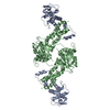
| ||||||||
| Unit cell |
|
-Components
| #1: Protein | / Cyclin A/CDK2-associated protein p19 / p19skp1 / p19A / RNA polymerase II elongation factor-like ...Cyclin A/CDK2-associated protein p19 / p19skp1 / p19A / RNA polymerase II elongation factor-like protein / Organ of Corti protein 2 / OCP-2 / OCP-II / Transcription elongation factor B / SIII Mass: 16827.127 Da / Num. of mol.: 1 Mutation: deletion: 38-43, 70-81; P2A; insertion: GGSG between 69 and 82 Source method: isolated from a genetically manipulated source Source: (gene. exp.) Homo sapiens (human) / Gene: EMC19, OCP2, SKP1, SKP1A, TCEB1L / Plasmid: pGEX / Production host:  Escherichia coli (E. coli) / Strain (production host): BL21(DE3) / References: UniProt: P63208 Escherichia coli (E. coli) / Strain (production host): BL21(DE3) / References: UniProt: P63208 |
|---|---|
| #2: Protein | Mass: 35848.684 Da / Num. of mol.: 1 / Fragment: Fbx4 residues 55-387 / Mutation: deletion: 150-170 Source method: isolated from a genetically manipulated source Source: (gene. exp.) Homo sapiens (human) / Gene: FBX4, FBXO4 / Plasmid: pGEX / Production host: Escherichia coli (E. coli) / Strain (production host): BL21(DE3) / References: UniProt: Q9UKT5 |
| #3: Water | ChemComp-HOH / Water Mass: 18.015 Da / Num. of mol.: 76 / Source method: isolated from a natural source / Formula: H2O Mass: 18.015 Da / Num. of mol.: 76 / Source method: isolated from a natural source / Formula: H2O |
-Experimental details
-Experiment
| Experiment | Method: X-RAY DIFFRACTION / Number of used crystals: 1 |
|---|
- Sample preparation
| Crystal | Density Matthews: 3.45 Å3/Da / Density % sol: 64.33 % |
|---|---|
| Crystal grow | Temperature: 277 K / Method: vapor diffusion, hanging drop / pH: 8 Details: 1.5-2 M sodium chloride, pH 8.0, VAPOR DIFFUSION, HANGING DROP, temperature 277.0K |
-Data collection
| Diffraction |
| ||||||||||||||||||
|---|---|---|---|---|---|---|---|---|---|---|---|---|---|---|---|---|---|---|---|
| Diffraction source |
| ||||||||||||||||||
| Detector |
| ||||||||||||||||||
| Radiation |
| ||||||||||||||||||
| Radiation wavelength |
| ||||||||||||||||||
| Reflection | Resolution: 2.8→50 Å / Num. all: 17477 / Num. obs: 17477 / % possible obs: 99.8 % / Observed criterion σ(F): 0 / Observed criterion σ(I): 0 / Redundancy: 14.3 % / Biso Wilson estimate: 84.1 Å2 / Rmerge(I) obs: 0.086 / Rsym value: 0.086 / Net I/σ(I): 31.5 | ||||||||||||||||||
| Reflection shell | Resolution: 2.8→2.9 Å / Redundancy: 14.3 % / Rmerge(I) obs: 0.856 / Mean I/σ(I) obs: 2.2 / Num. unique all: 1843 / Rsym value: 0.856 / % possible all: 100 |

- Processing
| Software |
| ||||||||||||||||||||||||||||||||||||||||||||||||||||||||||||||||||||||||||||||||||||||||||||||||||||||||||||||||||||||||||||||||||||||||||||||||||||||||||||||||||||||||||||||||||||||||||||||||||||||||
|---|---|---|---|---|---|---|---|---|---|---|---|---|---|---|---|---|---|---|---|---|---|---|---|---|---|---|---|---|---|---|---|---|---|---|---|---|---|---|---|---|---|---|---|---|---|---|---|---|---|---|---|---|---|---|---|---|---|---|---|---|---|---|---|---|---|---|---|---|---|---|---|---|---|---|---|---|---|---|---|---|---|---|---|---|---|---|---|---|---|---|---|---|---|---|---|---|---|---|---|---|---|---|---|---|---|---|---|---|---|---|---|---|---|---|---|---|---|---|---|---|---|---|---|---|---|---|---|---|---|---|---|---|---|---|---|---|---|---|---|---|---|---|---|---|---|---|---|---|---|---|---|---|---|---|---|---|---|---|---|---|---|---|---|---|---|---|---|---|---|---|---|---|---|---|---|---|---|---|---|---|---|---|---|---|---|---|---|---|---|---|---|---|---|---|---|---|---|---|---|---|---|
| Refinement | Method to determine structure: SAD / Resolution: 2.8→50 Å / Cor.coef. Fo:Fc: 0.938 / Cor.coef. Fo:Fc free: 0.912 / SU B: 38.305 / SU ML: 0.333 / TLS residual ADP flag: LIKELY RESIDUAL / Cross valid method: THROUGHOUT / σ(F): 0 / ESU R: 0.678 / ESU R Free: 0.372 Stereochemistry target values: MAXIMUM LIKELIHOOD WITH PHASES Details: HYDROGENS HAVE BEEN ADDED IN THE RIDING POSITIONS
| ||||||||||||||||||||||||||||||||||||||||||||||||||||||||||||||||||||||||||||||||||||||||||||||||||||||||||||||||||||||||||||||||||||||||||||||||||||||||||||||||||||||||||||||||||||||||||||||||||||||||
| Solvent computation | Ion probe radii: 0.8 Å / Shrinkage radii: 0.8 Å / VDW probe radii: 1.2 Å / Solvent model: MASK | ||||||||||||||||||||||||||||||||||||||||||||||||||||||||||||||||||||||||||||||||||||||||||||||||||||||||||||||||||||||||||||||||||||||||||||||||||||||||||||||||||||||||||||||||||||||||||||||||||||||||
| Displacement parameters | Biso mean: 37.752 Å2
| ||||||||||||||||||||||||||||||||||||||||||||||||||||||||||||||||||||||||||||||||||||||||||||||||||||||||||||||||||||||||||||||||||||||||||||||||||||||||||||||||||||||||||||||||||||||||||||||||||||||||
| Refinement step | Cycle: LAST / Resolution: 2.8→50 Å
| ||||||||||||||||||||||||||||||||||||||||||||||||||||||||||||||||||||||||||||||||||||||||||||||||||||||||||||||||||||||||||||||||||||||||||||||||||||||||||||||||||||||||||||||||||||||||||||||||||||||||
| Refine LS restraints |
| ||||||||||||||||||||||||||||||||||||||||||||||||||||||||||||||||||||||||||||||||||||||||||||||||||||||||||||||||||||||||||||||||||||||||||||||||||||||||||||||||||||||||||||||||||||||||||||||||||||||||
| LS refinement shell | Resolution: 2.8→2.873 Å / Total num. of bins used: 20
| ||||||||||||||||||||||||||||||||||||||||||||||||||||||||||||||||||||||||||||||||||||||||||||||||||||||||||||||||||||||||||||||||||||||||||||||||||||||||||||||||||||||||||||||||||||||||||||||||||||||||
| Refinement TLS params. | Method: refined / Refine-ID: X-RAY DIFFRACTION
| ||||||||||||||||||||||||||||||||||||||||||||||||||||||||||||||||||||||||||||||||||||||||||||||||||||||||||||||||||||||||||||||||||||||||||||||||||||||||||||||||||||||||||||||||||||||||||||||||||||||||
| Refinement TLS group |
|
