 Movie
Movie Controller


+ Open data

- Basic information
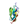
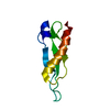
| Entry | Database: PDB / ID: 3l1x | ||||||
|---|---|---|---|---|---|---|---|
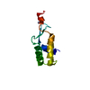
| Title | Crystal Structure of U-box Domain of Human E4B Ubiquitin Ligase | ||||||
 Components Components | Ubiquitin conjugation factor E4 B | ||||||
 Keywords Keywords |  LIGASE / E3 UBIQUITIN LIGASE / E4 UBIQUITIN LIGASE / U-BOX DOMAIN / Ubl conjugation pathway LIGASE / E3 UBIQUITIN LIGASE / E4 UBIQUITIN LIGASE / U-BOX DOMAIN / Ubl conjugation pathway | ||||||
| Function / homology |  Function and homology information Function and homology informationgranzyme-mediated apoptotic signaling pathway / ubiquitin-ubiquitin ligase activity / : / response to UV / ubiquitin ligase complex / RING-type E3 ubiquitin transferase / protein polyubiquitination / ubiquitin-dependent protein catabolic process / proteasome-mediated ubiquitin-dependent protein catabolic process / enzyme binding ...granzyme-mediated apoptotic signaling pathway / ubiquitin-ubiquitin ligase activity / : / response to UV / ubiquitin ligase complex / RING-type E3 ubiquitin transferase / protein polyubiquitination / ubiquitin-dependent protein catabolic process / proteasome-mediated ubiquitin-dependent protein catabolic process / enzyme binding / nucleus / cytoplasmSimilarity search - Function | ||||||
| Biological species |  Homo sapiens (human) Homo sapiens (human) | ||||||
| Method | X-RAY DIFFRACTION / SYNCHROTRON / MOLECULAR REPLACEMENT / Resolution: 2.6 Å | ||||||
 Authors Authors | Benirschke, R. / Thompson, J.R. / Mer, G. | ||||||
 Citation Citation | Journal: Structure / Year: 2010 Title: Molecular Basis for the Association of Human E4B U Box Ubiquitin Ligase with E2-Conjugating Enzymes UbcH5c and Ubc4. Authors: Benirschke, R.C. / Thompson, J.R. / Nomine, Y. / Wasielewski, E. / Juranic, N. / Macura, S. / Hatakeyama, S. / Nakayama, K.I. / Botuyan, M.V. / Mer, G. | ||||||
| History |
|
- Structure visualization
| Structure viewer | Molecule: MolmilJmol/JSmol |
|---|
- Downloads & links
-Download
| PDBx/mmCIF format | 3l1x.cif.gz | 45.3 KB | Display | PDBx/mmCIF format |
|---|---|---|---|---|
| PDB format | pdb3l1x.ent.gz | 31.5 KB | Display | PDB format |
| PDBx/mmJSON format | 3l1x.json.gz | Tree view | PDBx/mmJSON format | |
| Others |  Other downloads Other downloads |
-Validation report
| Arichive directory | https://data.pdbj.org/pub/pdb/validation_reports/l1/3l1xftp://data.pdbj.org/pub/pdb/validation_reports/l1/3l1x | HTTPS FTP |
|---|
-Related structure data
| Related structure data | 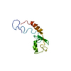 2kreC  3l1yC 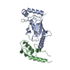 3l1zC  2ezj C: citing same article ( S: Starting model for refinement |
|---|---|
| Similar structure data |


-Links
 PDBj
PDBj
- Assembly
| Deposited unit | 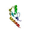
| ||||||||
|---|---|---|---|---|---|---|---|---|---|
| 1 |
| ||||||||
| Unit cell |
| ||||||||
| Components on special symmetry positions |
|
-Components
| #1: Protein | Mass: 11548.991 Da / Num. of mol.: 1 / Fragment: U box domain, residues 1208-1302 Source method: isolated from a genetically manipulated source Source: (gene. exp.) Homo sapiens (human) / Gene: UBE4B, HDNB1, KIAA0684, UFD2 / Plasmid: pET28b / Production host:  Escherichia coli (E. coli) / Strain (production host): BL21 (DE3) / References: UniProt: O95155 Escherichia coli (E. coli) / Strain (production host): BL21 (DE3) / References: UniProt: O95155 |
|---|---|
| #2: Water | ChemComp-HOH / Water Mass: 18.015 Da / Num. of mol.: 33 / Source method: isolated from a natural source / Formula: H2O Mass: 18.015 Da / Num. of mol.: 33 / Source method: isolated from a natural source / Formula: H2O |
-Experimental details
-Experiment
| Experiment | Method: X-RAY DIFFRACTION / Number of used crystals: 1 |
|---|
- Sample preparation
| Crystal | Density Matthews: 2.09 Å3/Da / Density % sol: 41.22 % |
|---|---|
| Crystal grow | Temperature: 295 K / Method: vapor diffusion, hanging drop Details: 2M TACSIMATE, VAPOR DIFFUSION, HANGING DROP, temperature 295K |
-Data collection
| Diffraction | Mean temperature: 100 K |
|---|---|
| Diffraction source | Source: SYNCHROTRON / Site: APS  / Beamline: 19-BM / Wavelength: 0.97918 Å / Beamline: 19-BM / Wavelength: 0.97918 Å |
| Detector | Type: ADSC QUANTUM 210r / Detector: CCD / Date: Feb 10, 2006 |
| Radiation | Protocol: SINGLE WAVELENGTH / Monochromatic (M) / Laue (L): M / Scattering type: x-ray |
| Radiation wavelength | Wavelength: 0.97918 Å / Relative weight: 1 |
| Reflection | Resolution: 2.6→24.074 Å / Num. obs: 3328 / % possible obs: 99.92 % / Redundancy: 37 % / Rmerge(I) obs: 0.139 / Net I/σ(I): 46.18 |
| Reflection shell | Resolution: 2.6→2.64 Å / Redundancy: 36.7 % / Rmerge(I) obs: 0.655 / Mean I/σ(I) obs: 7.29 / % possible all: 99 |
- Processing
| Software |
| ||||||||||||||||||||||||||||||||||||||||
|---|---|---|---|---|---|---|---|---|---|---|---|---|---|---|---|---|---|---|---|---|---|---|---|---|---|---|---|---|---|---|---|---|---|---|---|---|---|---|---|---|---|
| Refinement | Method to determine structure: MOLECULAR REPLACEMENT Starting model: 2EZJ 2ezj Resolution: 2.6→24.07 Å / SU ML: 0.34 / Isotropic thermal model: Isotropic / σ(F): 1.34 / Stereochemistry target values: ML
| ||||||||||||||||||||||||||||||||||||||||
| Solvent computation | Shrinkage radii: 0.9 Å / VDW probe radii: 1.11 Å / Solvent model: FLAT BULK SOLVENT MODEL / Bsol: 68.115 Å2 / ksol: 0.38 e/Å3 | ||||||||||||||||||||||||||||||||||||||||
| Displacement parameters | Biso mean: 42.58 Å2 | ||||||||||||||||||||||||||||||||||||||||
| Refinement step | Cycle: LAST / Resolution: 2.6→24.07 Å
| ||||||||||||||||||||||||||||||||||||||||
| Refine LS restraints |
| ||||||||||||||||||||||||||||||||||||||||
| LS refinement shell |
| ||||||||||||||||||||||||||||||||||||||||
| Refinement TLS params. | Method: refined / Origin x: -4.2179 Å / Origin y: 19.6556 Å / Origin z: 31.0241 Å
| ||||||||||||||||||||||||||||||||||||||||
| Refinement TLS group | Selection details: chain A |
