 Movie
Movie Controller

[English] 日本語
 Yorodumi
- PDB-3kyq: Lipid-induced Conformational Switch Controls Fusion Activity of L... -
+ Open data

- Basic information
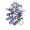
| Entry | Database: PDB / ID: 3kyq | ||||||
|---|---|---|---|---|---|---|---|
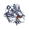
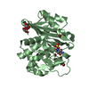
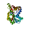
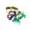
| Title | Lipid-induced Conformational Switch Controls Fusion Activity of Longin Domain SNARE Ykt6 | ||||||
 Components Components | Synaptobrevin homolog YKT6 | ||||||
 Keywords Keywords |  TRANSFERASE / v-SNARE homolog / lipid binding / Cytoplasmic vesicle / ER-Golgi transport / Golgi apparatus / Lipoprotein / Palmitate / Prenylation / Protein transport / Transport TRANSFERASE / v-SNARE homolog / lipid binding / Cytoplasmic vesicle / ER-Golgi transport / Golgi apparatus / Lipoprotein / Palmitate / Prenylation / Protein transport / Transport | ||||||
| Function / homology |  Function and homology information Function and homology informationIntra-Golgi traffic / protein-cysteine S-palmitoyltransferase activity / COPI-mediated anterograde transport / vesicle targeting / basal dendrite / COPII-mediated vesicle transport / SNARE complex / SNAP receptor activity / vesicle docking involved in exocytosis / apical dendrite ...Intra-Golgi traffic / protein-cysteine S-palmitoyltransferase activity / COPI-mediated anterograde transport / vesicle targeting / basal dendrite / COPII-mediated vesicle transport / SNARE complex / SNAP receptor activity / vesicle docking involved in exocytosis / apical dendrite / retrograde transport, endosome to Golgi / plasma membrane => GO:0005886 / endoplasmic reticulum to Golgi vesicle-mediated transport / Transferases; Acyltransferases; Transferring groups other than aminoacyl groups / cytoplasmic vesicle membrane / protein transport / endosome / Golgi membrane / neuronal cell body / Golgi apparatus / endoplasmic reticulum / mitochondrion / membrane / cytosol / cytoplasmSimilarity search - Function | ||||||
| Biological species |  Rattus norvegicus (Norway rat) Rattus norvegicus (Norway rat) | ||||||
| Method | X-RAY DIFFRACTION / MOLECULAR REPLACEMENT / Resolution: 2.443 Å | ||||||
 Authors Authors | Yu, J. / Wen, W.Y. / Zhang, M.J. | ||||||
 Citation Citation | Journal: Mol.Cell / Year: 2010 Title: Lipid-Induced Conformational Switch Controls Fusion Activity of Longin Domain SNARE Ykt6 Authors: Wen, W.Y. / Yu, J. / Pan, L.F. / Wei, Z.Y. / Weng, J.W. / Wang, W.N. / Ong, Y.S. / Tran, T.H.T. / Hong, W.J. / Zhang, M.J. | ||||||
| History |
|
- Structure visualization
| Structure viewer | Molecule: MolmilJmol/JSmol |
|---|
- Downloads & links
-Download
| PDBx/mmCIF format | 3kyq.cif.gz | 55.5 KB | Display | PDBx/mmCIF format |
|---|---|---|---|---|
| PDB format | pdb3kyq.ent.gz | 38.7 KB | Display | PDB format |
| PDBx/mmJSON format | 3kyq.json.gz | Tree view | PDBx/mmJSON format | |
| Others |  Other downloads Other downloads |
-Validation report
| Arichive directory | https://data.pdbj.org/pub/pdb/validation_reports/ky/3kyqftp://data.pdbj.org/pub/pdb/validation_reports/ky/3kyq | HTTPS FTP |
|---|
-Related structure data
| Related structure data |  3bw6S S: Starting model for refinement |
|---|---|
| Similar structure data |

-Links
 PDBj
PDBj

- Assembly
| Deposited unit | 
| ||||||||
|---|---|---|---|---|---|---|---|---|---|
| 1 |
| ||||||||
| Unit cell |
| ||||||||
| Components on special symmetry positions |
|
-Components
| #1: Protein | Mass: 22511.650 Da / Num. of mol.: 1 Source method: isolated from a genetically manipulated source Source: (gene. exp.) Rattus norvegicus (Norway rat) / Gene: Ykt6 / Plasmid: modified pET32a / Production host:  Escherichia coli (E. coli) / Strain (production host): BL21 DE3 Escherichia coli (E. coli) / Strain (production host): BL21 DE3References: UniProt: Q5EGY4, Transferases; Acyltransferases; Transferring groups other than aminoacyl groups | ||||||
|---|---|---|---|---|---|---|---|
| #2: Chemical | Sulfate  Mass: 96.063 Da / Num. of mol.: 2 / Source method: obtained synthetically / Formula: SO4 Mass: 96.063 Da / Num. of mol.: 2 / Source method: obtained synthetically / Formula: SO4#3: Chemical | ChemComp-DPV / | 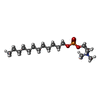  Mass: 351.462 Da / Num. of mol.: 1 / Source method: obtained synthetically / Formula: C17H38NO4P Mass: 351.462 Da / Num. of mol.: 1 / Source method: obtained synthetically / Formula: C17H38NO4P#4: Water | ChemComp-HOH / | Water Mass: 18.015 Da / Num. of mol.: 86 / Source method: isolated from a natural source / Formula: H2O Mass: 18.015 Da / Num. of mol.: 86 / Source method: isolated from a natural source / Formula: H2ONonpolymer details | THE LIGAND DPV (DODECYLPHO | |
-Experimental details
-Experiment
| Experiment | Method: X-RAY DIFFRACTION / Number of used crystals: 1 |
|---|
- Sample preparation
| Crystal | Density Matthews: 1.97 Å3/Da / Density % sol: 37.52 % |
|---|---|
| Crystal grow | Temperature: 289 K / Method: vapor diffusion, hanging drop / pH: 8.5 Details: 2M (NH4)2SO4, 0.1M Tris, pH 8.5, VAPOR DIFFUSION, HANGING DROP, temperature 289K |
-Data collection
| Diffraction | Mean temperature: 100 K |
|---|---|
| Diffraction source | Source: ROTATING ANODE / Type: RIGAKU MICROMAX-007 / Wavelength: 1.5418 Å |
| Detector | Type: RIGAKU RAXIS IV++ / Detector: IMAGE PLATE / Date: Jul 1, 2008 |
| Radiation | Monochromator: GRAPHITE / Protocol: SINGLE WAVELENGTH / Monochromatic (M) / Laue (L): M / Scattering type: x-ray |
| Radiation wavelength | Wavelength: 1.5418 Å / Relative weight: 1 |
| Reflection | Resolution: 2.443→30 Å / Num. all: 6900 / Num. obs: 6788 / % possible obs: 100 % / Redundancy: 5.1 % / Rmerge(I) obs: 0.067 / Net I/σ(I): 10 |
| Reflection shell | Resolution: 2.443→2.506 Å / Redundancy: 5.2 % / Rmerge(I) obs: 0.336 / Mean I/σ(I) obs: 2.2 / % possible all: 100 |
- Processing
| Software |
| ||||||||||||||||||||||||||||||||||||||||||||||||||||||||||||||||||||||||||||||||||||||||||
|---|---|---|---|---|---|---|---|---|---|---|---|---|---|---|---|---|---|---|---|---|---|---|---|---|---|---|---|---|---|---|---|---|---|---|---|---|---|---|---|---|---|---|---|---|---|---|---|---|---|---|---|---|---|---|---|---|---|---|---|---|---|---|---|---|---|---|---|---|---|---|---|---|---|---|---|---|---|---|---|---|---|---|---|---|---|---|---|---|---|---|---|
| Refinement | Method to determine structure: MOLECULAR REPLACEMENT Starting model: PDB ENTRY 3BW6 Resolution: 2.443→30 Å / Cor.coef. Fo:Fc: 0.937 / Cor.coef. Fo:Fc free: 0.885 / SU B: 18.161 / SU ML: 0.215 / TLS residual ADP flag: LIKELY RESIDUAL / Cross valid method: THROUGHOUT / ESU R Free: 0.342 / Stereochemistry target values: MAXIMUM LIKELIHOOD / Details: HYDROGENS HAVE BEEN ADDED IN THE RIDING POSITIONS
| ||||||||||||||||||||||||||||||||||||||||||||||||||||||||||||||||||||||||||||||||||||||||||
| Solvent computation | Ion probe radii: 0.8 Å / Shrinkage radii: 0.8 Å / VDW probe radii: 1.2 Å / Solvent model: MASK | ||||||||||||||||||||||||||||||||||||||||||||||||||||||||||||||||||||||||||||||||||||||||||
| Displacement parameters | Biso mean: 33.673 Å2
| ||||||||||||||||||||||||||||||||||||||||||||||||||||||||||||||||||||||||||||||||||||||||||
| Refinement step | Cycle: LAST / Resolution: 2.443→30 Å
| ||||||||||||||||||||||||||||||||||||||||||||||||||||||||||||||||||||||||||||||||||||||||||
| Refine LS restraints |
| ||||||||||||||||||||||||||||||||||||||||||||||||||||||||||||||||||||||||||||||||||||||||||
| LS refinement shell | Resolution: 2.443→2.506 Å / Total num. of bins used: 20
| ||||||||||||||||||||||||||||||||||||||||||||||||||||||||||||||||||||||||||||||||||||||||||
| Refinement TLS params. | Method: refined / Origin x: -9.4557 Å / Origin y: 6.7386 Å / Origin z: 14.9936 Å
|
