 Movie
Movie Controller

[English] 日本語
 Yorodumi
- PDB-3kwo: Crystal Structure of Putative Bacterioferritin from Campylobacter... -
+ Open data

- Basic information
| Entry | Database: PDB / ID: 3kwo | ||||||
|---|---|---|---|---|---|---|---|
| Title | Crystal Structure of Putative Bacterioferritin from Campylobacter jejuni | ||||||
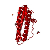
 Components Components | Putative bacterioferritin | ||||||
 Keywords Keywords | OXIDOREDUCTASE / alpha-helix / bacterial ferritin fold / Structural Genomics / Center for Structural Genomics of Infectious Diseases / CSGID / Cytoplasm / Iron / Iron storage / Metal-binding | ||||||
| Function / homology |  Function and homology informationOxidoreductases; Oxidizing metal ions / DNA protection / ferric iron binding / intracellular iron ion homeostasis / oxidoreductase activity / cytoplasm Function and homology informationOxidoreductases; Oxidizing metal ions / DNA protection / ferric iron binding / intracellular iron ion homeostasis / oxidoreductase activity / cytoplasmSimilarity search - Function | ||||||
| Biological species |   Campylobacter jejuni (Campylobacter) Campylobacter jejuni (Campylobacter) | ||||||
| Method | X-RAY DIFFRACTION / SYNCHROTRON / SAD / Resolution: 1.985 Å | ||||||
 Authors Authors | Kim, Y. / Gu, M. / Papazisi, L. / Anderson, W. / Joachimiak, A. / Center for Structural Genomics of Infectious Diseases (CSGID) | ||||||
 Citation Citation | Journal: To be Published Title: Crystal Structure of Putative Bacterioferritin from Campylobacter jejuni Authors: Kim, Y. / Gu, M. / Papazisi, L. / Anderson, W. / Joachimiak, A. | ||||||
| History |
|
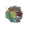
- Structure visualization
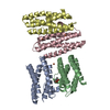
| Structure viewer | Molecule: MolmilJmol/JSmol |
|---|
- Downloads & links
-Download
| PDBx/mmCIF format | 3kwo.cif.gz | 266.7 KB | Display | PDBx/mmCIF format |
|---|---|---|---|---|
| PDB format | pdb3kwo.ent.gz | 226.3 KB | Display | PDB format |
| PDBx/mmJSON format | 3kwo.json.gz | Tree view | PDBx/mmJSON format | |
| Others |  Other downloads Other downloads |
-Validation report
| Arichive directory | https://data.pdbj.org/pub/pdb/validation_reports/kw/3kwoftp://data.pdbj.org/pub/pdb/validation_reports/kw/3kwo | HTTPS FTP |
|---|
-Related structure data
| Similar structure data | |
|---|---|
| Other databases |

-Links
 PDBj
PDBj


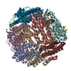
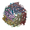
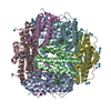
- Assembly
| Deposited unit | 
| ||||||||
|---|---|---|---|---|---|---|---|---|---|
| 1 | 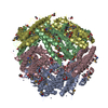
| ||||||||
| Unit cell |
| ||||||||
| Components on special symmetry positions |
|
-Components
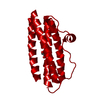
-Protein , 1 types, 4 molecules ABCD
| #1: Protein | Mass: 17780.426 Da / Num. of mol.: 4 Source method: isolated from a genetically manipulated source Source: (gene. exp.) Campylobacter jejuni (Campylobacter) / Species: subsp. jejuni NCTC 11168 / Strain: NCTC 11168 / Plasmid: Magic / Production host: Escherichia coli (E. coli) / Strain (production host): BL21 / References: UniProt: Q0P891 |
|---|
-Non-polymers , 5 types, 628 molecules 


| #2: Chemical | ChemComp-ACY / Acetic acid Mass: 60.052 Da / Num. of mol.: 24 / Source method: obtained synthetically / Formula: C2H4O2 Mass: 60.052 Da / Num. of mol.: 24 / Source method: obtained synthetically / Formula: C2H4O2#3: Chemical | Glycerol Mass: 92.094 Da / Num. of mol.: 2 / Source method: obtained synthetically / Formula: C3H8O3 Mass: 92.094 Da / Num. of mol.: 2 / Source method: obtained synthetically / Formula: C3H8O3#4: Chemical | ChemComp-ZN /  Mass: 65.409 Da / Num. of mol.: 26 / Source method: obtained synthetically / Formula: Zn Mass: 65.409 Da / Num. of mol.: 26 / Source method: obtained synthetically / Formula: Zn#5: Chemical | 1,4-Butanediol Mass: 90.121 Da / Num. of mol.: 3 / Source method: obtained synthetically / Formula: C4H10O2 Mass: 90.121 Da / Num. of mol.: 3 / Source method: obtained synthetically / Formula: C4H10O2#6: Water | ChemComp-HOH / | WaterMass: 18.015 Da / Num. of mol.: 573 / Source method: isolated from a natural source / Formula: H2O |
|---|
-Experimental details
-Experiment
| Experiment | Method: X-RAY DIFFRACTION / Number of used crystals: 1 |
|---|
- Sample preparation
| Crystal | Density Matthews: 2.34 Å3/Da / Density % sol: 47.42 % |
|---|---|
| Crystal grow | Temperature: 289 K / Method: vapor diffusion, sitting drop / pH: 8 Details: 0.2 M Zinc acetate, 0.1 M Imidazole pH 8.0, 20% (v/v) 1,4-bdiolutaiediol, VAPOR DIFFUSION, SITTING DROP, temperature 289K |
-Data collection
| Diffraction | Mean temperature: 100 K |
|---|---|
| Diffraction source | Source: SYNCHROTRON / Site: APS  / Beamline: 19-BM / Wavelength: 0.97934 Å / Beamline: 19-BM / Wavelength: 0.97934 Å |
| Detector | Type: ADSC QUANTUM 210r / Detector: CCD / Date: Jul 19, 2009 / Details: mirrors |
| Radiation | Monochromator: double crystal monochromator / Protocol: SAD / Monochromatic (M) / Laue (L): M / Scattering type: x-ray |
| Radiation wavelength | Wavelength: 0.97934 Å / Relative weight: 1 |
| Reflection | Resolution: 1.98→28.78 Å / Num. all: 43678 / Num. obs: 43678 / % possible obs: 98.5 % / Observed criterion σ(F): 0 / Observed criterion σ(I): 0 / Redundancy: 6.3 % / Biso Wilson estimate: 21.3 Å2 / Rmerge(I) obs: 0.135 / Net I/σ(I): 9.54 |
| Reflection shell | Resolution: 1.98→2.01 Å / Redundancy: 4.8 % / Mean I/σ(I) obs: 2.51 / Rsym value: 0.854 / % possible all: 76.3 |
- Processing
| Software |
| ||||||||||||||||||||||||||||||||||||||||||||||||||||||||||||||||||||||||||||||||||||||||||||||||||||||||||||||||||||||||||||||||||
|---|---|---|---|---|---|---|---|---|---|---|---|---|---|---|---|---|---|---|---|---|---|---|---|---|---|---|---|---|---|---|---|---|---|---|---|---|---|---|---|---|---|---|---|---|---|---|---|---|---|---|---|---|---|---|---|---|---|---|---|---|---|---|---|---|---|---|---|---|---|---|---|---|---|---|---|---|---|---|---|---|---|---|---|---|---|---|---|---|---|---|---|---|---|---|---|---|---|---|---|---|---|---|---|---|---|---|---|---|---|---|---|---|---|---|---|---|---|---|---|---|---|---|---|---|---|---|---|---|---|---|---|
| Refinement | Method to determine structure: SAD / Resolution: 1.985→28.78 Å / SU ML: 0.21 / Isotropic thermal model: mixed / σ(F): 1.96 / Phase error: 18.53 / Stereochemistry target values: MLHL
| ||||||||||||||||||||||||||||||||||||||||||||||||||||||||||||||||||||||||||||||||||||||||||||||||||||||||||||||||||||||||||||||||||
| Solvent computation | Shrinkage radii: 0.9 Å / VDW probe radii: 1.11 Å / Solvent model: FLAT BULK SOLVENT MODEL / Bsol: 41.044 Å2 / ksol: 0.343 e/Å3 | ||||||||||||||||||||||||||||||||||||||||||||||||||||||||||||||||||||||||||||||||||||||||||||||||||||||||||||||||||||||||||||||||||
| Displacement parameters |
| ||||||||||||||||||||||||||||||||||||||||||||||||||||||||||||||||||||||||||||||||||||||||||||||||||||||||||||||||||||||||||||||||||
| Refinement step | Cycle: LAST / Resolution: 1.985→28.78 Å
| ||||||||||||||||||||||||||||||||||||||||||||||||||||||||||||||||||||||||||||||||||||||||||||||||||||||||||||||||||||||||||||||||||
| Refine LS restraints |
| ||||||||||||||||||||||||||||||||||||||||||||||||||||||||||||||||||||||||||||||||||||||||||||||||||||||||||||||||||||||||||||||||||
| LS refinement shell |
| ||||||||||||||||||||||||||||||||||||||||||||||||||||||||||||||||||||||||||||||||||||||||||||||||||||||||||||||||||||||||||||||||||
| Refinement TLS params. | Refine-ID: X-RAY DIFFRACTION
| ||||||||||||||||||||||||||||||||||||||||||||||||||||||||||||||||||||||||||||||||||||||||||||||||||||||||||||||||||||||||||||||||||
| Refinement TLS group |
|
