 Movie
Movie Controller


+ Open data

- Basic information
| Entry | Database: PDB / ID: 3kuq | ||||||
|---|---|---|---|---|---|---|---|
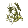
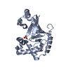
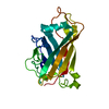
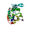
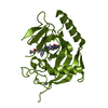
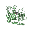
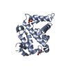
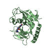
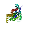
| Title | Crystal structure of the DLC1 RhoGAP domain | ||||||
 Components Components | Rho GTPase-activating protein 7 | ||||||
 Keywords Keywords | HYDROLASE ACTIVATOR /  Structural Genomics Consortium / GTPase activation / SGC / Alternative splicing / Cytoplasm / Phosphoprotein / Polymorphism Structural Genomics Consortium / GTPase activation / SGC / Alternative splicing / Cytoplasm / Phosphoprotein / Polymorphism | ||||||
| Function / homology |  Function and homology information Function and homology informationhindbrain morphogenesis / negative regulation of focal adhesion assembly / regulation of Rho protein signal transduction / focal adhesion assembly / regulation of small GTPase mediated signal transduction / negative regulation of stress fiber assembly / negative regulation of Rho protein signal transduction / cortical actin cytoskeleton / RHOB GTPase cycle / positive regulation of execution phase of apoptosis ...hindbrain morphogenesis / negative regulation of focal adhesion assembly / regulation of Rho protein signal transduction / focal adhesion assembly / regulation of small GTPase mediated signal transduction / negative regulation of stress fiber assembly / negative regulation of Rho protein signal transduction / cortical actin cytoskeleton / RHOB GTPase cycle / positive regulation of execution phase of apoptosis / RHOC GTPase cycle / RHOQ GTPase cycle / CDC42 GTPase cycle / RHOA GTPase cycle / heart morphogenesis / positive regulation of protein dephosphorylation / forebrain development / RAC1 GTPase cycle / SH2 domain binding / GTPase activator activity / negative regulation of cell migration / neural tube closure / caveola / regulation of actin cytoskeleton organization / ruffle membrane / activation of cysteine-type endopeptidase activity involved in apoptotic process / regulation of cell shape / actin cytoskeleton organization / membrane raft / negative regulation of cell population proliferation / intracellular membrane-bounded organelle / focal adhesion / lipid binding / apoptotic process / endoplasmic reticulum / signal transduction / nucleus / cytosol / cytoplasmSimilarity search - Function | ||||||
| Biological species |  Homo sapiens (human) Homo sapiens (human) | ||||||
| Method | X-RAY DIFFRACTION / SYNCHROTRON / MOLECULAR REPLACEMENT / molecular replacement / Resolution: 2.3 Å | ||||||
 Authors Authors | Nedyalkova, L. / Tempel, W. / Tong, Y. / MacKenzie, F. / Shen, L. / Zhong, N. / Arrowsmith, C.H. / Edwards, A.M. / Bountra, C. / Weigelt, J. ...Nedyalkova, L. / Tempel, W. / Tong, Y. / MacKenzie, F. / Shen, L. / Zhong, N. / Arrowsmith, C.H. / Edwards, A.M. / Bountra, C. / Weigelt, J. / Bochkarev, A. / Park, H. / Structural Genomics Consortium (SGC) | ||||||
 Citation Citation | Journal: to be published Title: Crystal structure of the DLC1 RhoGAP domain Authors: Nedyalkova, L. / Tempel, W. / Tong, Y. / MacKenzie, F. / Shen, L. / Zhong, N. / Arrowsmith, C.H. / Edwards, A.M. / Bountra, C. / Weigelt, J. / Bochkarev, A. / Park, H. | ||||||
| History |
|
- Structure visualization
| Structure viewer | Molecule: MolmilJmol/JSmol |
|---|
- Downloads & links
-Download
| PDBx/mmCIF format | 3kuq.cif.gz | 52.2 KB | Display | PDBx/mmCIF format |
|---|---|---|---|---|
| PDB format | pdb3kuq.ent.gz | 36.1 KB | Display | PDB format |
| PDBx/mmJSON format | 3kuq.json.gz | Tree view | PDBx/mmJSON format | |
| Others |  Other downloads Other downloads |
-Validation report
| Arichive directory | https://data.pdbj.org/pub/pdb/validation_reports/ku/3kuqftp://data.pdbj.org/pub/pdb/validation_reports/ku/3kuq | HTTPS FTP |
|---|
-Related structure data
| Related structure data |  3fk2S S: Starting model for refinement |
|---|---|
| Similar structure data |

-Links
 PDBj
PDBj


- Assembly
| Deposited unit | 
| ||||||||
|---|---|---|---|---|---|---|---|---|---|
| 1 |
| ||||||||
| Unit cell |
| ||||||||
| Components on special symmetry positions |
|
-Components
| #1: Protein | Mass: 26038.930 Da / Num. of mol.: 1 / Fragment: Rho-GAP domain Source method: isolated from a genetically manipulated source Source: (gene. exp.) Homo sapiens (human) / Gene: DLC1, ARHGAP7, KIAA1723, STARD12 / Plasmid: pET28-MHL / Production host:  Escherichia coli (E. coli) / Strain (production host): BL21-V2R-pRARE2 / References: UniProt: Q96QB1 Escherichia coli (E. coli) / Strain (production host): BL21-V2R-pRARE2 / References: UniProt: Q96QB1 | ||
|---|---|---|---|
| #2: Chemical | ChemComp-UNX /   Num. of mol.: 4 / Source method: obtained synthetically Num. of mol.: 4 / Source method: obtained synthetically#3: Water | ChemComp-HOH / | Water Mass: 18.015 Da / Num. of mol.: 30 / Source method: isolated from a natural source / Formula: H2O Mass: 18.015 Da / Num. of mol.: 30 / Source method: isolated from a natural source / Formula: H2O |
-Experimental details
-Experiment
| Experiment | Method: X-RAY DIFFRACTION / Number of used crystals: 1 |
|---|
- Sample preparation
| Crystal | Density Matthews: 2.62 Å3/Da / Density % sol: 53.14 % |
|---|---|
| Crystal grow | Temperature: 291 K / pH: 7.5 Details: 30% PEG-3350, 0.2M sodium chloride, 5% glycerol, 0.02M HEPES, pH 7.5, vapor diffusion, hanging drop, temperature 291K, VAPOR DIFFUSION, HANGING DROP |
-Data collection
| Diffraction | Mean temperature: 100 K |
|---|---|
| Diffraction source | Source: SYNCHROTRON / Site: APS  / Beamline: 19-ID / Wavelength: 0.97915 / Beamline: 19-ID / Wavelength: 0.97915 |
| Detector | Type: ADSC Q315 / Detector: CCD / Date: Nov 5, 2009 |
| Radiation | Protocol: SINGLE WAVELENGTH / Scattering type: x-ray |
| Radiation wavelength | Wavelength: 0.97915 Å / Relative weight: 1 |
| Reflection | Resolution: 2.3→50 Å / Num. obs: 12307 / % possible obs: 98.2 % |
| Reflection shell | Resolution: 2.3→2.34 Å / Rmerge(I) obs: 0.685 / % possible all: 87.5 |
-Phasing
| Phasing | Method: molecular replacement |
|---|
- Processing
| Software |
| ||||||||||||||||||||||||||||||||||||||||||||||||||||||||||||||||||||||||||||||||||||||||||||||||||||||||||||||||||||||||||||||||||||||||||||||||||||||||||||||||||||||||||
|---|---|---|---|---|---|---|---|---|---|---|---|---|---|---|---|---|---|---|---|---|---|---|---|---|---|---|---|---|---|---|---|---|---|---|---|---|---|---|---|---|---|---|---|---|---|---|---|---|---|---|---|---|---|---|---|---|---|---|---|---|---|---|---|---|---|---|---|---|---|---|---|---|---|---|---|---|---|---|---|---|---|---|---|---|---|---|---|---|---|---|---|---|---|---|---|---|---|---|---|---|---|---|---|---|---|---|---|---|---|---|---|---|---|---|---|---|---|---|---|---|---|---|---|---|---|---|---|---|---|---|---|---|---|---|---|---|---|---|---|---|---|---|---|---|---|---|---|---|---|---|---|---|---|---|---|---|---|---|---|---|---|---|---|---|---|---|---|---|---|---|---|
| Refinement | Method to determine structure: MOLECULAR REPLACEMENT Starting model: pdb entry 3fk2 Resolution: 2.3→48.75 Å / Cor.coef. Fo:Fc: 0.925 / Cor.coef. Fo:Fc free: 0.914 / SU B: 6.188 / SU ML: 0.15 / Cross valid method: THROUGHOUT / ESU R: 0.269 / ESU R Free: 0.222 / Stereochemistry target values: MAXIMUM LIKELIHOOD Details: HYDROGENS HAVE BEEN ADDED IN THE RIDING POSITIONS U VALUES : REFINED INDIVIDUALLY. The programs CHAINSAW, PHENIX, ARP/WARP, COOT and MOLPROBITY were also used during refinement.
| ||||||||||||||||||||||||||||||||||||||||||||||||||||||||||||||||||||||||||||||||||||||||||||||||||||||||||||||||||||||||||||||||||||||||||||||||||||||||||||||||||||||||||
| Solvent computation | Ion probe radii: 0.8 Å / Shrinkage radii: 0.8 Å / VDW probe radii: 1.4 Å / Solvent model: MASK | ||||||||||||||||||||||||||||||||||||||||||||||||||||||||||||||||||||||||||||||||||||||||||||||||||||||||||||||||||||||||||||||||||||||||||||||||||||||||||||||||||||||||||
| Displacement parameters | Biso mean: 20.67 Å2
| ||||||||||||||||||||||||||||||||||||||||||||||||||||||||||||||||||||||||||||||||||||||||||||||||||||||||||||||||||||||||||||||||||||||||||||||||||||||||||||||||||||||||||
| Refinement step | Cycle: LAST / Resolution: 2.3→48.75 Å
| ||||||||||||||||||||||||||||||||||||||||||||||||||||||||||||||||||||||||||||||||||||||||||||||||||||||||||||||||||||||||||||||||||||||||||||||||||||||||||||||||||||||||||
| Refine LS restraints |
| ||||||||||||||||||||||||||||||||||||||||||||||||||||||||||||||||||||||||||||||||||||||||||||||||||||||||||||||||||||||||||||||||||||||||||||||||||||||||||||||||||||||||||
| LS refinement shell | Resolution: 2.3→2.36 Å / Total num. of bins used: 20
|
