 Movie
Movie Controller

[English] 日本語
 Yorodumi
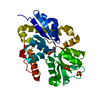
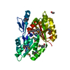
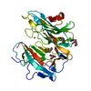
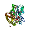
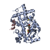
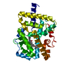
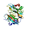
- PDB-3ksj: The alkanesulfonate-binding protein SsuA from Xabthomonas axonopo... -
+ Open data

- Basic information
| Entry | Database: PDB / ID: 3ksj | ||||||
|---|---|---|---|---|---|---|---|
| Title | The alkanesulfonate-binding protein SsuA from Xabthomonas axonopodis pv. citri bound to MES | ||||||
 Components Components | Nitrate transport protein | ||||||
 Keywords Keywords |  TRANSPORT PROTEIN / SsuA / alkanesulfonates / periplasmic-binding protein TRANSPORT PROTEIN / SsuA / alkanesulfonates / periplasmic-binding protein | ||||||
| Function / homology |  Function and homology information Function and homology informationATPase-coupled transmembrane transporter activity / periplasmic space / membraneSimilarity search - Function | ||||||
| Biological species |  Xanthomonas axonopodis pv. citri (bacteria) Xanthomonas axonopodis pv. citri (bacteria) | ||||||
| Method | X-RAY DIFFRACTION / SYNCHROTRON / MOLECULAR REPLACEMENT / molecular replacement / Resolution: 2 Å | ||||||
 Authors Authors | Balan, A. / Araujo, F.T. / Barbosa, J.A.R.G. | ||||||
 Citation Citation | Journal: To be Published Title: The crystallographic structure of the SsuA protein reveals how alkanesulfonates enter into the cell Authors: Balan, A. / Araujo, F.T. / Barbosa, J.A.R.G. | ||||||
| History |
|
- Structure visualization
| Structure viewer | Molecule: MolmilJmol/JSmol |
|---|
- Downloads & links
-Download
| PDBx/mmCIF format | 3ksj.cif.gz | 72.3 KB | Display | PDBx/mmCIF format |
|---|---|---|---|---|
| PDB format | pdb3ksj.ent.gz | 51.7 KB | Display | PDB format |
| PDBx/mmJSON format | 3ksj.json.gz | Tree view | PDBx/mmJSON format | |
| Others |  Other downloads Other downloads |
-Validation report
| Arichive directory | https://data.pdbj.org/pub/pdb/validation_reports/ks/3ksjftp://data.pdbj.org/pub/pdb/validation_reports/ks/3ksj | HTTPS FTP |
|---|
-Related structure data
| Related structure data |  3ksxC 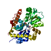 3e4rS S: Starting model for refinement C: citing same article ( |
|---|---|
| Similar structure data |


-Links
 PDBj
PDBj- Assembly
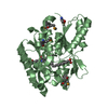
| Deposited unit | 
| ||||||||
|---|---|---|---|---|---|---|---|---|---|
| 1 |
| ||||||||
| Unit cell |
|
-Components
| #1: Protein | Mass: 34566.688 Da / Num. of mol.: 1 Source method: isolated from a genetically manipulated source Source: (gene. exp.) Xanthomonas axonopodis pv. citri (bacteria)Strain: 306 / Gene: ssuA / Plasmid: pET28a / Production host: Escherichia coli (E. coli) / Strain (production host): BL21(DE3) / References: UniProt: Q8PHQ1 |
|---|---|
| #2: Chemical | ChemComp-MES / MES (buffer)  Mass: 195.237 Da / Num. of mol.: 1 / Source method: obtained synthetically / Formula: C6H13NO4S / Comment: pH buffer*YM Mass: 195.237 Da / Num. of mol.: 1 / Source method: obtained synthetically / Formula: C6H13NO4S / Comment: pH buffer*YM |
| #3: Water | ChemComp-HOH / Water Mass: 18.015 Da / Num. of mol.: 106 / Source method: isolated from a natural source / Formula: H2O Mass: 18.015 Da / Num. of mol.: 106 / Source method: isolated from a natural source / Formula: H2O |
-Experimental details
-Experiment
| Experiment | Method: X-RAY DIFFRACTION / Number of used crystals: 1 |
|---|
- Sample preparation
| Crystal | Density Matthews: 1.78 Å3/Da / Density % sol: 30.75 % |
|---|---|
| Crystal grow | Temperature: 298 K / Method: vapor diffusion, hanging drop / pH: 6.5 Details: 0.2 M MES, pH 6.5, 0.1 M NaCl, 1.6 M ammonium sulfate, VAPOR DIFFUSION, HANGING DROP, temperature 298K |
-Data collection
| Diffraction | Mean temperature: 298 K |
|---|---|
| Diffraction source | Source: SYNCHROTRON / Site: LNLS  / Beamline: W01B-MX2 / Wavelength: 1.46 Å / Beamline: W01B-MX2 / Wavelength: 1.46 Å |
| Detector | Type: MARMOSAIC 225 mm CCD / Detector: CCD / Date: Oct 8, 2009 / Details: mirrors |
| Radiation | Monochromator: GRAPHITE / Protocol: SINGLE WAVELENGTH / Monochromatic (M) / Laue (L): M / Scattering type: x-ray |
| Radiation wavelength | Wavelength: 1.46 Å / Relative weight: 1 |
| Reflection | Resolution: 2→30 Å / Num. all: 16418 / Num. obs: 15613 / % possible obs: 95.1 % / Observed criterion σ(F): 2 / Observed criterion σ(I): 2 / Redundancy: 3.4 % / Biso Wilson estimate: 22.7 Å2 / Rmerge(I) obs: 0.113 / Χ2: 1.1 / Net I/σ(I): 6.7 |
| Reflection shell | Resolution: 2→2.07 Å / Redundancy: 2.9 % / Rmerge(I) obs: 0.402 / Num. unique all: 1321 / Χ2: 0.892 / % possible all: 80.5 |
-Phasing
| Phasing | Method: molecular replacement | |||||||||
|---|---|---|---|---|---|---|---|---|---|---|
| Phasing MR |
|
- Processing
| Software |
| |||||||||||||||||||||||||||||||||||||||||||||||||||||||||||||||||
|---|---|---|---|---|---|---|---|---|---|---|---|---|---|---|---|---|---|---|---|---|---|---|---|---|---|---|---|---|---|---|---|---|---|---|---|---|---|---|---|---|---|---|---|---|---|---|---|---|---|---|---|---|---|---|---|---|---|---|---|---|---|---|---|---|---|---|
| Refinement | Method to determine structure: MOLECULAR REPLACEMENT Starting model: PDB ENTRY 3E4R Resolution: 2→24.09 Å / Cor.coef. Fo:Fc: 0.955 / Cor.coef. Fo:Fc free: 0.91 / WRfactor Rfree: 0.247 / WRfactor Rwork: 0.174 / Occupancy max: 1 / Occupancy min: 0.5 / FOM work R set: 0.809 / SU B: 5.413 / SU ML: 0.15 / SU R Cruickshank DPI: 0.248 / SU Rfree: 0.21 / Isotropic thermal model: Overall / Cross valid method: THROUGHOUT / σ(F): 0 / σ(I): 2 / ESU R: 0.248 / ESU R Free: 0.21 / Stereochemistry target values: MAXIMUM LIKELIHOOD Details: HYDROGENS HAVE BEEN ADDED IN THE RIDING POSITIONS U VALUES : REFINED INDIVIDUALLY
| |||||||||||||||||||||||||||||||||||||||||||||||||||||||||||||||||
| Solvent computation | Ion probe radii: 0.8 Å / Shrinkage radii: 0.8 Å / VDW probe radii: 1.4 Å / Solvent model: MASK | |||||||||||||||||||||||||||||||||||||||||||||||||||||||||||||||||
| Displacement parameters | Biso max: 163.32 Å2 / Biso mean: 27.435 Å2 / Biso min: 9.25 Å2
| |||||||||||||||||||||||||||||||||||||||||||||||||||||||||||||||||
| Refinement step | Cycle: LAST / Resolution: 2→24.09 Å
| |||||||||||||||||||||||||||||||||||||||||||||||||||||||||||||||||
| Refine LS restraints |
| |||||||||||||||||||||||||||||||||||||||||||||||||||||||||||||||||
| LS refinement shell | Resolution: 2→2.049 Å / Total num. of bins used: 20
|
