[English] 日本語
Yorodumi
- PDB-3kks: Crystal structure of catalytic core domain of BIV integrase in cr... -

+
Open data


ID or keywords:

Loading...

-
Basic information

Entry
Database: PDB / ID: 3kks
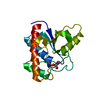
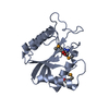
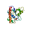
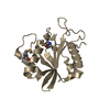
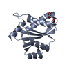
TitleCrystal structure of catalytic core domain of BIV integrase in crystal form II
ComponentsIntegrase
KeywordsDNA BINDING PROTEIN / beta-strands flanked by alpha-helices
Function / homology
Function and homology information


microtubule-dependent intracellular transport of viral material towards nucleus / Hydrolases; Acting on peptide bonds (peptidases); Aspartic endopeptidases / host cell / retroviral ribonuclease H / exoribonuclease H / exoribonuclease H activity / DNA integration / RNA-directed DNA polymerase / viral genome integration into host DNA / establishment of integrated proviral latency ...microtubule-dependent intracellular transport of viral material towards nucleus / Hydrolases; Acting on peptide bonds (peptidases); Aspartic endopeptidases / host cell / retroviral ribonuclease H / exoribonuclease H / exoribonuclease H activity / DNA integration / RNA-directed DNA polymerase / viral genome integration into host DNA / establishment of integrated proviral latency / RNA-directed DNA polymerase activity / RNA-DNA hybrid ribonuclease activity / Transferases; Transferring phosphorus-containing groups; Nucleotidyltransferases / viral nucleocapsid / DNA recombination / structural constituent of virion / Hydrolases; Acting on ester bonds / DNA-directed DNA polymerase / aspartic-type endopeptidase activity / DNA-directed DNA polymerase activity / symbiont entry into host cell / proteolysis / DNA binding / RNA binding / zinc ion binding
Similarity search - Function
Ribonuclease H-like superfamily/Ribonuclease H / Reverse transcriptase thumb / Reverse transcriptase thumb domain / Integrase Zinc binding domain / Zinc finger integrase-type profile. / Integrase-like, N-terminal / Integrase DNA binding domain / Integrase, C-terminal domain superfamily, retroviral / Integrase, N-terminal zinc-binding domain / Integrase, C-terminal, retroviral ...Ribonuclease H-like superfamily/Ribonuclease H / Reverse transcriptase thumb / Reverse transcriptase thumb domain / Integrase Zinc binding domain / Zinc finger integrase-type profile. / Integrase-like, N-terminal / Integrase DNA binding domain / Integrase, C-terminal domain superfamily, retroviral / Integrase, N-terminal zinc-binding domain / Integrase, C-terminal, retroviral / Integrase DNA binding domain profile. / RNase H / Integrase core domain / Integrase, catalytic core / Integrase catalytic domain profile. / Retroviral nucleocapsid Gag protein p24, C-terminal domain / Gag protein p24 C-terminal domain / Ribonuclease H domain / RNase H type-1 domain profile. / Reverse transcriptase (RNA-dependent DNA polymerase) / Reverse transcriptase domain / Reverse transcriptase (RT) catalytic domain profile. / Retropepsins / Retroviral aspartyl protease / Aspartyl protease, retroviral-type family profile. / Peptidase A2A, retrovirus, catalytic / Retrovirus capsid, C-terminal / Retrovirus capsid, N-terminal / zinc finger / Zinc knuckle / Zinc finger, CCHC-type superfamily / Zinc finger, CCHC-type / Zinc finger CCHC-type profile. / Nucleotidyltransferase; domain 5 / Aspartic peptidase, active site / Eukaryotic and viral aspartyl proteases active site. / Ribonuclease H superfamily / Aspartic peptidase domain superfamily / Ribonuclease H-like superfamily / Reverse transcriptase/Diguanylate cyclase domain / DNA/RNA polymerase superfamily / 2-Layer Sandwich / Alpha Beta
Similarity search - Domain/homology
ACETATE ION / Gag-Pol polyprotein
Similarity search - Component
Biological speciesBovine immunodeficiency virus
MethodX-RAY DIFFRACTION / MOLECULAR REPLACEMENT / Resolution: 2.2 Å
AuthorsShen, Y.
CitationJournal: Protein Cell / Year: 2010
Title: Crystal structures of catalytic core domain of BIV integrase: implications for the interaction between integrase and target DNA
Authors: Yao, X. / Fang, S. / Qiao, W. / Geng, Y. / Shen, Y.
History
DepositionNov 6, 2009Deposition site: RCSB / Processing site: PDBJ
Revision 1.0Sep 22, 2010Provider: repository / Type: Initial release
Revision 1.1Jul 13, 2011Group: Source and taxonomy / Version format compliance
Revision 1.2Mar 28, 2012Group: Database references
Revision 1.3Nov 1, 2023Group: Data collection / Database references ...Data collection / Database references / Derived calculations / Refinement description
Category: chem_comp_atom / chem_comp_bond ...chem_comp_atom / chem_comp_bond / database_2 / pdbx_initial_refinement_model / struct_ref_seq_dif / struct_site
Item: _database_2.pdbx_DOI / _database_2.pdbx_database_accession ..._database_2.pdbx_DOI / _database_2.pdbx_database_accession / _struct_ref_seq_dif.details / _struct_site.pdbx_auth_asym_id / _struct_site.pdbx_auth_comp_id / _struct_site.pdbx_auth_seq_id

-
Structure visualization

Structure viewerMolecule:
MolmilJmol/JSmol

Downloads & links

-
Assembly

Deposited unit
A: Integrase
B: Integrase
hetero molecules


Theoretical massNumber of molelcules
Total (without water)34,4516
Polymers34,1492
Non-polymers3024
Water1,67593
1
A: Integrase
hetero molecules


Theoretical massNumber of molelcules
Total (without water)17,2253
Polymers17,0741
Non-polymers1512
Water181
TypeNameSymmetry operationNumber
identity operation1_555x,y,z1
2
B: Integrase
hetero molecules


Theoretical massNumber of molelcules
Total (without water)17,2253
Polymers17,0741
Non-polymers1512
Water181
TypeNameSymmetry operationNumber
identity operation1_555x,y,z1
3
A: Integrase
hetero molecules

A: Integrase
hetero molecules


Theoretical massNumber of molelcules
Total (without water)34,4516
Polymers34,1492
Non-polymers3024
Water362
TypeNameSymmetry operationNumber
identity operation1_555x,y,z1
crystal symmetry operation8_555-y,-x,-z+1/21
Buried area3700 Å2
ΔGint-11 kcal/mol
Surface area12550 Å2
MethodPISA
4
B: Integrase
hetero molecules

B: Integrase
hetero molecules


Theoretical massNumber of molelcules
Total (without water)34,4516
Polymers34,1492
Non-polymers3024
Water362
TypeNameSymmetry operationNumber
identity operation1_555x,y,z1
crystal symmetry operation7_555y,x,-z1
Buried area3600 Å2
ΔGint-14 kcal/mol
Surface area12630 Å2
MethodPISA
Unit cell
Length a, b, c (Å)88.054, 88.054, 81.775
Angle α, β, γ (deg.)90.00, 90.00, 90.00
Int Tables number92
Space group name H-MP41212
Components on special symmetry positions
IDModelComponents
11A-12-

HOH

-
Components

#1: Protein Integrase / / IN


Mass: 17074.330 Da / Num. of mol.: 2 / Fragment: catalytic core domain
Source method: isolated from a genetically manipulated source
Source: (gene. exp.) Bovine immunodeficiency virus / Strain: R29 / Gene: gag-pol / Production host: Escherichia coli (E. coli) / References: UniProt: P19560
#2: Chemical ChemComp-ACT / ACETATE ION / Acetate


Mass: 59.044 Da / Num. of mol.: 2 / Source method: obtained synthetically / Formula: C2H3O2
#3: Chemical ChemComp-GOL / GLYCEROL / GLYCERIN / PROPANE-1,2,3-TRIOL / Glycerol


Mass: 92.094 Da / Num. of mol.: 2 / Source method: obtained synthetically / Formula: C3H8O3
#4: Water ChemComp-HOH / water / Water


Mass: 18.015 Da / Num. of mol.: 93 / Source method: isolated from a natural source / Formula: H2O

-
Experimental details

-
Experiment

ExperimentMethod: X-RAY DIFFRACTION / Number of used crystals: 1

-
Sample preparation

CrystalDensity Matthews: 2.32 Å3/Da / Density % sol: 47 %
Crystal growTemperature: 293 K / Method: vapor diffusion, sitting drop / pH: 6.5
Details: PEG 10000, sodium acetate, pH 6.5, VAPOR DIFFUSION, SITTING DROP, temperature 293K

-
Data collection

DiffractionMean temperature: 200 K
Diffraction sourceSource: ROTATING ANODE / Type: RIGAKU MICROMAX-007 / Wavelength: 1.5418 Å
DetectorType: RIGAKU RAXIS HTC / Detector: IMAGE PLATE / Date: Apr 1, 2009
RadiationMonochromator: Mirror / Protocol: SINGLE WAVELENGTH / Monochromatic (M) / Laue (L): M / Scattering type: x-ray
Radiation wavelengthWavelength: 1.5418 Å / Relative weight: 1
ReflectionResolution: 2.2→50 Å / Num. obs: 16868 / % possible obs: 99.8 % / Observed criterion σ(F): 2 / Observed criterion σ(I): 2 / Redundancy: 12.7 % / Biso Wilson estimate: 27 Å2 / Rsym value: 0.103 / Net I/σ(I): 26.5
Reflection shellResolution: 2.2→2.28 Å / Redundancy: 13 % / Mean I/σ(I) obs: 6.6 / Num. unique all: 1643 / Rsym value: 0.5 / % possible all: 99.9

-
Processing

Software
NameVersionClassification
HKL-2000data collection
PHASERphasing
CNS1.2refinement
HKL-2000data reduction
HKL-2000data scaling
RefinementMethod to determine structure: MOLECULAR REPLACEMENT
Starting model: PDB ID 3KKR
Resolution: 2.2→19.48 Å / Rfactor Rfree error: 0.01 / Data cutoff high absF: 154327.63 / Data cutoff low absF: 0 / Isotropic thermal model: RESTRAINED / Cross valid method: THROUGHOUT / σ(F): 0 / Stereochemistry target values: Engh & Huber / Details: BULK SOLVENT MODEL USED
RfactorNum. reflection% reflectionSelection details
Rfree0.272 797 4.9 %RANDOM
Rwork0.239 ---
obs0.239 16303 96.7 %-
Solvent computationSolvent model: FLAT MODEL / Bsol: 46.3327 Å2 / ksol: 0.4 e/Å3
Displacement parametersBiso mean: 34 Å2
Baniso -1Baniso -2Baniso -3
1-4.17 Å20 Å20 Å2
2--4.17 Å20 Å2
3----8.35 Å2
Refine analyze
FreeObs
Luzzati coordinate error0.34 Å0.3 Å
Luzzati d res low-5 Å
Luzzati sigma a0.25 Å0.21 Å
Refinement stepCycle: LAST / Resolution: 2.2→19.48 Å
ProteinNucleic acidLigandSolventTotal
Num. atoms2359 0 20 93 2472
Refine LS restraints
Refine-IDTypeDev idealDev ideal target
X-RAY DIFFRACTIONc_bond_d0.006
X-RAY DIFFRACTIONc_angle_deg1.3
X-RAY DIFFRACTIONc_dihedral_angle_d22.7
X-RAY DIFFRACTIONc_improper_angle_d0.78
X-RAY DIFFRACTIONc_mcbond_it1.331.5
X-RAY DIFFRACTIONc_mcangle_it2.132
X-RAY DIFFRACTIONc_scbond_it2.152
X-RAY DIFFRACTIONc_scangle_it3.192.5
LS refinement shellResolution: 2.2→2.34 Å / Rfactor Rfree error: 0.025 / Total num. of bins used: 6
RfactorNum. reflection% reflection
Rfree0.281 123 4.8 %
Rwork0.269 2427 -
obs--92.6 %
Xplor file
Refine-IDSerial noParam fileTopol file
X-RAY DIFFRACTION1protein_rep.paramprotein.top
X-RAY DIFFRACTION2water_rep.paramwater_rep.top
X-RAY DIFFRACTION3gol.paramgol.top
X-RAY DIFFRACTION4act.paramact.top

+
About Yorodumi

-
News

-
Feb 9, 2022. New format data for meta-information of EMDB entries

New format data for meta-information of EMDB entries

  • Version 3 of the EMDB header file is now the official format.
  • The previous official version 1.9 will be removed from the archive.

Related info.:EMDB header

External links:wwPDB to switch to version 3 of the EMDB data model

-
Aug 12, 2020. Covid-19 info

Covid-19 info

URL: https://pdbj.org/emnavi/covid19.php

New page: Covid-19 featured information page in EM Navigator.

Related info.:Covid-19 info / Mar 5, 2020. Novel coronavirus structure data

+
Mar 5, 2020. Novel coronavirus structure data

Novel coronavirus structure data

Related info.:Yorodumi Speices / Aug 12, 2020. Covid-19 info

External links:COVID-19 featured content - PDBj / Molecule of the Month (242):Coronavirus Proteases

+
Jan 31, 2019. EMDB accession codes are about to change! (news from PDBe EMDB page)

EMDB accession codes are about to change! (news from PDBe EMDB page)

  • The allocation of 4 digits for EMDB accession codes will soon come to an end. Whilst these codes will remain in use, new EMDB accession codes will include an additional digit and will expand incrementally as the available range of codes is exhausted. The current 4-digit format prefixed with “EMD-” (i.e. EMD-XXXX) will advance to a 5-digit format (i.e. EMD-XXXXX), and so on. It is currently estimated that the 4-digit codes will be depleted around Spring 2019, at which point the 5-digit format will come into force.
  • The EM Navigator/Yorodumi systems omit the EMD- prefix.

Related info.:Q: What is EMD? / ID/Accession-code notation in Yorodumi/EM Navigator

External links:EMDB Accession Codes are Changing Soon! / Contact to PDBj

+
Jul 12, 2017. Major update of PDB

Major update of PDB

  • wwPDB released updated PDB data conforming to the new PDBx/mmCIF dictionary.
  • This is a major update changing the version number from 4 to 5, and with Remediation, in which all the entries are updated.
  • In this update, many items about electron microscopy experimental information are reorganized (e.g. em_software).
  • Now, EM Navigator and Yorodumi are based on the updated data.

External links:wwPDB Remediation / Enriched Model Files Conforming to OneDep Data Standards Now Available in the PDB FTP Archive

-
Yorodumi

Thousand views of thousand structures

  • Yorodumi is a browser for structure data from EMDB, PDB, SASBDB, etc.
  • This page is also the successor to EM Navigator detail page, and also detail information page/front-end page for Omokage search.
  • The word "yorodu" (or yorozu) is an old Japanese word meaning "ten thousand". "mi" (miru) is to see.

Related info.:EMDB / PDB / SASBDB / Comparison of 3 databanks / Yorodumi Search / Aug 31, 2016. New EM Navigator & Yorodumi / Yorodumi Papers / Jmol/JSmol / Function and homology information / Changes in new EM Navigator and Yorodumi

Read more