 Movie
Movie Controller

[English] 日本語
 Yorodumi
- PDB-3kg1: Crystal structure of SnoaB, a cofactor-independent oxygenase from... -
+ Open data

- Basic information
| Entry | Database: PDB / ID: 3kg1 | ||||||
|---|---|---|---|---|---|---|---|
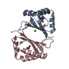
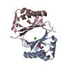
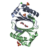
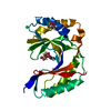
| Title | Crystal structure of SnoaB, a cofactor-independent oxygenase from Streptomyces nogalater, mutant N63A | ||||||
 Components Components | SnoaB | ||||||
 Keywords Keywords |  OXIDOREDUCTASE / polyketide / anthracycline / oxygenase / cofactor-independent OXIDOREDUCTASE / polyketide / anthracycline / oxygenase / cofactor-independent | ||||||
| Function / homology |  Function and homology information Function and homology informationdeoxynogalonate monooxygenase / oxidoreductase activity, acting on single donors with incorporation of molecular oxygen, incorporation of one atom of oxygen (internal monooxygenases or internal mixed function oxidases) / antibiotic biosynthetic process Similarity search - Function | ||||||
| Biological species |  Streptomyces nogalater (bacteria) Streptomyces nogalater (bacteria) | ||||||
| Method | X-RAY DIFFRACTION / SYNCHROTRON / MOLECULAR REPLACEMENT / molecular replacement / Resolution: 2.5 Å | ||||||
 Authors Authors | Koskiniemi, H. / Grocholski, T. / Lindqvist, Y. / Mantsala, P. / Niemi, J. / Schneider, G. | ||||||
 Citation Citation | Journal: Biochemistry / Year: 2010 Title: Crystal structure of the cofactor-independent monooxygenase SnoaB from Streptomyces nogalater: implications for the reaction mechanism Authors: Grocholski, T. / Koskiniemi, H. / Lindqvist, Y. / Mantsala, P. / Niemi, J. / Schneider, G. #1: Journal: Acta Crystallogr.,Sect.F / Year: 2009 Title: Expression, purification and crystallization of the cofactor-independent monooxygenase SnoaB from the nogalamycin biosynthetic pathway Authors: Koskiniemi, H. / Grocholski, T. / Schneider, G. / Niemi, J. | ||||||
| History |
|
- Structure visualization
| Structure viewer | Molecule: MolmilJmol/JSmol |
|---|
- Downloads & links
-Download
| PDBx/mmCIF format | 3kg1.cif.gz | 72.2 KB | Display | PDBx/mmCIF format |
|---|---|---|---|---|
| PDB format | pdb3kg1.ent.gz | 53.7 KB | Display | PDB format |
| PDBx/mmJSON format | 3kg1.json.gz | Tree view | PDBx/mmJSON format | |
| Others |  Other downloads Other downloads |
-Validation report
| Arichive directory | https://data.pdbj.org/pub/pdb/validation_reports/kg/3kg1ftp://data.pdbj.org/pub/pdb/validation_reports/kg/3kg1 | HTTPS FTP |
|---|
-Related structure data


-Links
 PDBj
PDBj- Assembly
| Deposited unit | 
| ||||||||||||||||||||||||||||||||||||||||||||||||||||||||||||||||||||||||||||||||||
|---|---|---|---|---|---|---|---|---|---|---|---|---|---|---|---|---|---|---|---|---|---|---|---|---|---|---|---|---|---|---|---|---|---|---|---|---|---|---|---|---|---|---|---|---|---|---|---|---|---|---|---|---|---|---|---|---|---|---|---|---|---|---|---|---|---|---|---|---|---|---|---|---|---|---|---|---|---|---|---|---|---|---|---|
| 1 | 
| ||||||||||||||||||||||||||||||||||||||||||||||||||||||||||||||||||||||||||||||||||
| 2 | 
| ||||||||||||||||||||||||||||||||||||||||||||||||||||||||||||||||||||||||||||||||||
| 3 | 
| ||||||||||||||||||||||||||||||||||||||||||||||||||||||||||||||||||||||||||||||||||
| Unit cell |
| ||||||||||||||||||||||||||||||||||||||||||||||||||||||||||||||||||||||||||||||||||
| Noncrystallographic symmetry (NCS) | NCS domain:
NCS domain segments: Ens-ID: 1 / Refine code: 4
|
-Components
| #1: Protein | Mass: 14311.804 Da / Num. of mol.: 3 / Fragment: oxygenase / Mutation: N63A Source method: isolated from a genetically manipulated source Source: (gene. exp.) Streptomyces nogalater (bacteria) / Gene: SnoaB / Plasmid: modified from pBAD/HisB / Production host: Escherichia coli (E. coli) / Strain (production host): TOP10 / References: UniProt: O54259#2: Chemical | Chloride  Mass: 35.453 Da / Num. of mol.: 2 / Source method: obtained synthetically / Formula: Cl Mass: 35.453 Da / Num. of mol.: 2 / Source method: obtained synthetically / Formula: Cl#3: Water | ChemComp-HOH / | Water Mass: 18.015 Da / Num. of mol.: 34 / Source method: isolated from a natural source / Formula: H2O Mass: 18.015 Da / Num. of mol.: 34 / Source method: isolated from a natural source / Formula: H2OSequence details | AUTHORS STATE THAT G25A IS IN FACT THE NATIVE SEQUENCE. THEY CONFIRMED THEIR NATIVE SEQUENCE BY DNA- ...AUTHORS STATE THAT G25A IS IN FACT THE NATIVE SEQUENCE. THEY CONFIRMED THEIR NATIVE SEQUENCE BY DNA-SEQUENCING | |
|---|
-Experimental details
-Experiment
| Experiment | Method: X-RAY DIFFRACTION / Number of used crystals: 1 |
|---|
- Sample preparation
| Crystal | Density Matthews: 1.98 Å3/Da / Density % sol: 31.94 % |
|---|---|
| Crystal grow | Temperature: 277 K / Method: vapor diffusion, sitting drop / pH: 8 Details: PEG MME 2K, KBr, pentaerythritol ethoxylate , pH 8.0, VAPOR DIFFUSION, SITTING DROP, temperature 277.0K |
-Data collection
| Diffraction | Mean temperature: 100 K |
|---|---|
| Diffraction source | Source: SYNCHROTRON / Site: ESRF  / Beamline: ID14-2 / Wavelength: 0.933 Å / Beamline: ID14-2 / Wavelength: 0.933 Å |
| Detector | Type: ADSC QUANTUM 4 / Detector: CCD / Date: Feb 12, 2009 |
| Radiation | Monochromator: Diamond (111), Ge(220) / Protocol: SINGLE WAVELENGTH / Scattering type: x-ray |
| Radiation wavelength | Wavelength: 0.933 Å / Relative weight: 1 |
| Reflection | Resolution: 2.5→30 Å / Num. all: 11301 / Num. obs: 11301 / % possible obs: 99.8 % / Observed criterion σ(F): 0 / Observed criterion σ(I): 0 / Redundancy: 6 % / Biso Wilson estimate: 52.5 Å2 / Rsym value: 0.103 / Net I/σ(I): 13.2 |
| Reflection shell | Resolution: 2.5→2.64 Å / Redundancy: 3.7 % / Mean I/σ(I) obs: 2 / Num. unique all: 1591 / Rsym value: 0.571 / % possible all: 99.3 |
-Phasing
| Phasing | Method: molecular replacement |
|---|
- Processing
| Software |
| ||||||||||||||||||||||||||||||||||||||||||||||||||||||||||||||||||||||||||||||||||||||||||||||||||||
|---|---|---|---|---|---|---|---|---|---|---|---|---|---|---|---|---|---|---|---|---|---|---|---|---|---|---|---|---|---|---|---|---|---|---|---|---|---|---|---|---|---|---|---|---|---|---|---|---|---|---|---|---|---|---|---|---|---|---|---|---|---|---|---|---|---|---|---|---|---|---|---|---|---|---|---|---|---|---|---|---|---|---|---|---|---|---|---|---|---|---|---|---|---|---|---|---|---|---|---|---|---|
| Refinement | Method to determine structure: MOLECULAR REPLACEMENT / Resolution: 2.5→29.42 Å / Cor.coef. Fo:Fc: 0.943 / Cor.coef. Fo:Fc free: 0.904 / Occupancy max: 1 / Occupancy min: 0 / SU B: 25.621 / SU ML: 0.261 / TLS residual ADP flag: LIKELY RESIDUAL / Cross valid method: THROUGHOUT / σ(F): 0 / ESU R: 0.71 / ESU R Free: 0.312 / Stereochemistry target values: MAXIMUM LIKELIHOOD
| ||||||||||||||||||||||||||||||||||||||||||||||||||||||||||||||||||||||||||||||||||||||||||||||||||||
| Solvent computation | Ion probe radii: 0.8 Å / Shrinkage radii: 0.8 Å / VDW probe radii: 1.4 Å / Solvent model: BABINET MODEL WITH MASK | ||||||||||||||||||||||||||||||||||||||||||||||||||||||||||||||||||||||||||||||||||||||||||||||||||||
| Displacement parameters | Biso max: 70.71 Å2 / Biso mean: 35.709 Å2 / Biso min: 17.85 Å2
| ||||||||||||||||||||||||||||||||||||||||||||||||||||||||||||||||||||||||||||||||||||||||||||||||||||
| Refinement step | Cycle: LAST / Resolution: 2.5→29.42 Å
| ||||||||||||||||||||||||||||||||||||||||||||||||||||||||||||||||||||||||||||||||||||||||||||||||||||
| Refine LS restraints |
| ||||||||||||||||||||||||||||||||||||||||||||||||||||||||||||||||||||||||||||||||||||||||||||||||||||
| Refine LS restraints NCS | Dom-ID: 1 / Ens-ID: 1 / Number: 1100 / Refine-ID: X-RAY DIFFRACTION
| ||||||||||||||||||||||||||||||||||||||||||||||||||||||||||||||||||||||||||||||||||||||||||||||||||||
| LS refinement shell | Resolution: 2.5→2.564 Å / Total num. of bins used: 20
| ||||||||||||||||||||||||||||||||||||||||||||||||||||||||||||||||||||||||||||||||||||||||||||||||||||
| Refinement TLS params. | Method: refined / Refine-ID: X-RAY DIFFRACTION
| ||||||||||||||||||||||||||||||||||||||||||||||||||||||||||||||||||||||||||||||||||||||||||||||||||||
| Refinement TLS group |
|
