cathepsin V / negative regulation of peptidase activity / peptidase inhibitor complex / Formation of the cornified envelope / peptide cross-linking / cornified envelope / RUNX1 regulates transcription of genes involved in differentiation of keratinocytes / Trafficking and processing of endosomal TLR / Assembly of collagen fibrils and other multimeric structures / cysteine-type endopeptidase inhibitor activity ...cathepsin V / negative regulation of peptidase activity / peptidase inhibitor complex / Formation of the cornified envelope / peptide cross-linking / cornified envelope / RUNX1 regulates transcription of genes involved in differentiation of keratinocytes / Trafficking and processing of endosomal TLR / Assembly of collagen fibrils and other multimeric structures / cysteine-type endopeptidase inhibitor activity / Activation of Matrix Metalloproteinases / cysteine-type endopeptidase activator activity involved in apoptotic process / extracellular matrix disassembly / keratinocyte differentiation / cysteine-type peptidase activity / MHC class II antigen presentation / Degradation of the extracellular matrix / lysosomal lumen / proteolysis involved in protein catabolic process / positive regulation of apoptotic signaling pathway / negative regulation of proteolysis / Endosomal/Vacuolar pathway / cell-cell adhesion / antigen processing and presentation of exogenous peptide antigen via MHC class II / protease binding / immune response / cysteine-type endopeptidase activity / serine-type endopeptidase activity / extracellular space / extracellular region / nucleoplasm / cytosol / cytoplasm Similarity search - Function
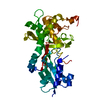
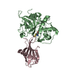
Proteinase inhibitor I25A, stefin / Proteinase inhibitor I25, cystatin, conserved site / Cysteine proteases inhibitors signature. / Cystatin domain / Cystatin-like domain / Cystatin domain / Nuclear Transport Factor 2; Chain: A, - #10 / Cystatin superfamily / Cathepsin propeptide inhibitor domain (I29) / Cathepsin propeptide inhibitor domain (I29) ...Proteinase inhibitor I25A, stefin / Proteinase inhibitor I25, cystatin, conserved site / Cysteine proteases inhibitors signature. / Cystatin domain / Cystatin-like domain / Cystatin domain / Nuclear Transport Factor 2; Chain: A, - #10 / Cystatin superfamily / Cathepsin propeptide inhibitor domain (I29) / Cathepsin propeptide inhibitor domain (I29) / Cathepsin propeptide inhibitor domain (I29) / Papain-like cysteine endopeptidase / : / Cysteine peptidase, asparagine active site / Eukaryotic thiol (cysteine) proteases asparagine active site. / Cysteine peptidase, histidine active site / Eukaryotic thiol (cysteine) proteases histidine active site. / Peptidase C1A, papain C-terminal / Papain family cysteine protease / Papain family cysteine protease / Cysteine proteinases / Cysteine peptidase, cysteine active site / Eukaryotic thiol (cysteine) proteases cysteine active site. / Cathepsin B; Chain A / Nuclear Transport Factor 2; Chain: A, / Papain-like cysteine peptidase superfamily / Roll / Alpha-Beta Complex / Alpha Beta Similarity search - Domain/homology
Mass: 18.015 Da / Num. of mol.: 722 / Source method: isolated from a natural source / Formula: H2O
Sequence details
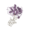
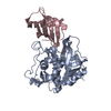
CATHEPSIN V WAS BLOCKED WITH MMTS (METHYL METHANETHIOSULFONATE), LEAVING -S-CH3 ATOMS ON ACTIVE ...CATHEPSIN V WAS BLOCKED WITH MMTS (METHYL METHANETHIOSULFONATE), LEAVING -S-CH3 ATOMS ON ACTIVE SITE CYSTEINE RESIDUE.
-
Experimental details
-
Experiment
Experiment
Method: X-RAY DIFFRACTION / Number of used crystals: 1
-
Sample preparation
Crystal
Density Matthews: 2.63 Å3/Da / Density % sol: 53.19 % / Mosaicity: 0.546 °
Crystal grow
Temperature: 293 K / Method: vapor diffusion, sitting drop / pH: 8 Details: 0.1M Tris-HCl, 12% PEG3000, pH 8.0, VAPOR DIFFUSION, SITTING DROP, temperature 293K
Resolution: 1.99→19.49 Å / Cor.coef. Fo:Fc: 0.961 / Cor.coef. Fo:Fc free: 0.941 / WRfactor Rfree: 0.205 / WRfactor Rwork: 0.164 / Occupancy max: 1 / Occupancy min: 0 / FOM work R set: 0.867 / SU R Cruickshank DPI: 0.166 / SU Rfree: 0.151 / Cross valid method: THROUGHOUT / σ(F): 0 / ESU R: 0.166 / ESU R Free: 0.151 / Stereochemistry target values: MAXIMUM LIKELIHOOD Details: HYDROGENS HAVE BEEN ADDED IN THE RIDING POSITIONS U VALUES: REFINED INDIVIDUALLY. The structure was refined also with MAIN
Rfactor
Num. reflection
% reflection
Selection details
Rfree
0.219
2586
5.1 %
RANDOM
Rwork
0.176
-
-
-
all
0.182
51180
-
-
obs
0.178
50986
99.62 %
-
Solvent computation
Ion probe radii: 0.8 Å / Shrinkage radii: 0.8 Å / VDW probe radii: 1.4 Å / Solvent model: MASK
In the structure databanks used in Yorodumi, some data are registered as the other names, "COVID-19 virus" and "2019-nCoV". Here are the details of the virus and the list of structure data.
Jan 31, 2019. EMDB accession codes are about to change! (news from PDBe EMDB page)
EMDB accession codes are about to change! (news from PDBe EMDB page)
The allocation of 4 digits for EMDB accession codes will soon come to an end. Whilst these codes will remain in use, new EMDB accession codes will include an additional digit and will expand incrementally as the available range of codes is exhausted. The current 4-digit format prefixed with “EMD-” (i.e. EMD-XXXX) will advance to a 5-digit format (i.e. EMD-XXXXX), and so on. It is currently estimated that the 4-digit codes will be depleted around Spring 2019, at which point the 5-digit format will come into force.
The EM Navigator/Yorodumi systems omit the EMD- prefix.
Related info.:Q: What is EMD? / ID/Accession-code notation in Yorodumi/EM Navigator
Yorodumi is a browser for structure data from EMDB, PDB, SASBDB, etc.
This page is also the successor to EM Navigator detail page, and also detail information page/front-end page for Omokage search.
The word "yorodu" (or yorozu) is an old Japanese word meaning "ten thousand". "mi" (miru) is to see.
Related info.:EMDB / PDB / SASBDB / Comparison of 3 databanks / Yorodumi Search / Aug 31, 2016. New EM Navigator & Yorodumi / Yorodumi Papers / Jmol/JSmol / Function and homology information / Changes in new EM Navigator and Yorodumi
 Movie
Movie Controller


 Open data
 Basic information
 Components
 Keywords
 cathepsin / protease-inhibitor complex /
  Function and homology information
 Authors
 Citation
 Structure visualization
 Downloads & links
 Other downloads


 PDBj
PDBj


 Assembly


 Mass: 18.015 Da / Num. of mol.: 722 / Source method: isolated from a natural source / Formula: H2O
 Sample preparation
 / Beamline: 5.2R / Wavelength: 1 Å
 Processing