 Movie
Movie Controller

[English] 日本語
 Yorodumi
- PDB-3ke4: Crystal structure of a PduO-Type ATP:Cob(I)alamin adenosyltransfe... -
+ Open data

- Basic information
| Entry | Database: PDB / ID: 3ke4 | ||||||
|---|---|---|---|---|---|---|---|
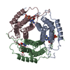
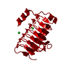
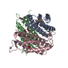
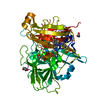
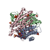
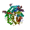
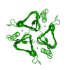
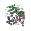
| Title | Crystal structure of a PduO-Type ATP:Cob(I)alamin adenosyltransferase from Bacillus cereus | ||||||
 Components Components | Hypothetical Cytosolic Protein | ||||||
 Keywords Keywords |  TRANSFERASE / Helix bundle TRANSFERASE / Helix bundle | ||||||
| Function / homology |  Function and homology information Function and homology informationcorrinoid adenosyltransferase / corrinoid adenosyltransferase activity / cobalamin biosynthetic process / ATP bindingSimilarity search - Function | ||||||
| Biological species |  Bacillus cereus (bacteria) Bacillus cereus (bacteria) | ||||||
| Method | X-RAY DIFFRACTION / SYNCHROTRON / MOLECULAR REPLACEMENT / Resolution: 1.9 Å | ||||||
 Authors Authors | Park, A.K. / Moon, J.H. / Chi, Y.M. | ||||||
 Citation Citation | Journal: To be Published Title: Crystal structure of a PduO-Type ATP:Cob(I)alamin adenosyltransferase from Bacillus cereus Authors: Park, A.K. / Moon, J.H. / Chi, Y.M. | ||||||
| History |
|
- Structure visualization
| Structure viewer | Molecule: MolmilJmol/JSmol |
|---|
- Downloads & links
-Download
| PDBx/mmCIF format | 3ke4.cif.gz | 121.3 KB | Display | PDBx/mmCIF format |
|---|---|---|---|---|
| PDB format | pdb3ke4.ent.gz | 93.7 KB | Display | PDB format |
| PDBx/mmJSON format | 3ke4.json.gz | Tree view | PDBx/mmJSON format | |
| Others |  Other downloads Other downloads |
-Validation report
| Arichive directory | https://data.pdbj.org/pub/pdb/validation_reports/ke/3ke4ftp://data.pdbj.org/pub/pdb/validation_reports/ke/3ke4 | HTTPS FTP |
|---|
-Related structure data
| Related structure data |  2ah6S S: Starting model for refinement |
|---|---|
| Similar structure data |


-Links
 PDBj
PDBj- Assembly
| Deposited unit | 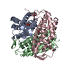
| ||||||||
|---|---|---|---|---|---|---|---|---|---|
| 1 |
| ||||||||
| Unit cell |
|
-Components
| #1: Protein | Mass: 23936.320 Da / Num. of mol.: 3 Source method: isolated from a genetically manipulated source Source: (gene. exp.) Bacillus cereus (bacteria) / Strain: ATCC 14579 / Plasmid: pET 28a / Production host: Escherichia coli (E. coli) / Strain (production host): BL21(DE3) / References: UniProt: Q813T7, corrinoid adenosyltransferase#2: Chemical | ChemComp-DIO / 1,4-Dioxane  Mass: 88.105 Da / Num. of mol.: 8 / Source method: obtained synthetically / Formula: C4H8O2 Mass: 88.105 Da / Num. of mol.: 8 / Source method: obtained synthetically / Formula: C4H8O2#3: Water | ChemComp-HOH / | Water Mass: 18.015 Da / Num. of mol.: 359 / Source method: isolated from a natural source / Formula: H2O Mass: 18.015 Da / Num. of mol.: 359 / Source method: isolated from a natural source / Formula: H2O |
|---|
-Experimental details
-Experiment
| Experiment | Method: X-RAY DIFFRACTION / Number of used crystals: 1 |
|---|
- Sample preparation
| Crystal | Density Matthews: 2.46 Å3/Da / Density % sol: 49.93 % / Mosaicity: 0.224 ° |
|---|---|
| Crystal grow | Temperature: 295 K / Method: vapor diffusion, hanging drop / pH: 6.5 Details: 0.1M MES pH 6.5, 1.52M ammonium sulfate, 9%(v/v) dioxane, VAPOR DIFFUSION, HANGING DROP, temperature 295K |
-Data collection
| Diffraction | Mean temperature: 77 K | ||||||||||||||||||||||||||||||||||||||||||||||||||||||||||||||||||
|---|---|---|---|---|---|---|---|---|---|---|---|---|---|---|---|---|---|---|---|---|---|---|---|---|---|---|---|---|---|---|---|---|---|---|---|---|---|---|---|---|---|---|---|---|---|---|---|---|---|---|---|---|---|---|---|---|---|---|---|---|---|---|---|---|---|---|---|
| Diffraction source | Source: SYNCHROTRON / Site: PAL/PLS  / Beamline: 6C1 / Wavelength: 1.23986 Å / Beamline: 6C1 / Wavelength: 1.23986 Å | ||||||||||||||||||||||||||||||||||||||||||||||||||||||||||||||||||
| Detector | Type: ADSC QUANTUM 210 / Detector: CCD / Date: Jan 29, 2008 | ||||||||||||||||||||||||||||||||||||||||||||||||||||||||||||||||||
| Radiation | Monochromator: Graphite / Protocol: SINGLE WAVELENGTH / Monochromatic (M) / Laue (L): M / Scattering type: x-ray | ||||||||||||||||||||||||||||||||||||||||||||||||||||||||||||||||||
| Radiation wavelength | Wavelength: 1.23986 Å / Relative weight: 1 | ||||||||||||||||||||||||||||||||||||||||||||||||||||||||||||||||||
| Reflection | Resolution: 1.9→50 Å / Num. obs: 53001 / % possible obs: 94.4 % / Redundancy: 3.9 % / Rmerge(I) obs: 0.04 / Χ2: 0.801 / Net I/σ(I): 14.2 | ||||||||||||||||||||||||||||||||||||||||||||||||||||||||||||||||||
| Reflection shell |
|
- Processing
| Software |
| ||||||||||||||||||||||||||||||||||||
|---|---|---|---|---|---|---|---|---|---|---|---|---|---|---|---|---|---|---|---|---|---|---|---|---|---|---|---|---|---|---|---|---|---|---|---|---|---|
| Refinement | Method to determine structure: MOLECULAR REPLACEMENT Starting model: PDB ENTRY 2ah6 Resolution: 1.9→39.64 Å / Occupancy max: 1 / Occupancy min: 0.5 / σ(F): 0
| ||||||||||||||||||||||||||||||||||||
| Solvent computation | Bsol: 58.322 Å2 | ||||||||||||||||||||||||||||||||||||
| Displacement parameters | Biso max: 82.4 Å2 / Biso mean: 24.478 Å2 / Biso min: 2.82 Å2
| ||||||||||||||||||||||||||||||||||||
| Refinement step | Cycle: LAST / Resolution: 1.9→39.64 Å
| ||||||||||||||||||||||||||||||||||||
| Refine LS restraints |
| ||||||||||||||||||||||||||||||||||||
| Xplor file |
|
