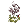
 Movie
Movie Controller


+ Open data

- Basic information
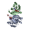
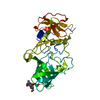
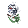
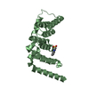
| Entry | Database: PDB / ID: 3kbf | ||||||
|---|---|---|---|---|---|---|---|
| Title | C. elegans Cu,Zn Superoxide Dismutase | ||||||
 Components Components | Superoxide dismutase [Cu-Zn] | ||||||
 Keywords Keywords |  OXIDOREDUCTASE / cu-zn superoxide dismutase / antioxidant / nematode / Disulfide bond / Metal-binding OXIDOREDUCTASE / cu-zn superoxide dismutase / antioxidant / nematode / Disulfide bond / Metal-binding | ||||||
| Function / homology |  Function and homology information Function and homology informationDetoxification of Reactive Oxygen Species / Platelet degranulation / regulation of vulval development / regulation of brood size / superoxide metabolic process / superoxide dismutase / superoxide dismutase activity / removal of superoxide radicals / copper ion binding / protein homodimerization activity ...Detoxification of Reactive Oxygen Species / Platelet degranulation / regulation of vulval development / regulation of brood size / superoxide metabolic process / superoxide dismutase / superoxide dismutase activity / removal of superoxide radicals / copper ion binding / protein homodimerization activity / mitochondrion / cytosol / cytoplasmSimilarity search - Function | ||||||
| Biological species |  Caenorhabditis elegans (invertebrata) Caenorhabditis elegans (invertebrata) | ||||||
| Method | X-RAY DIFFRACTION / SYNCHROTRON / MOLECULAR REPLACEMENT / Resolution: 1.3 Å | ||||||
 Authors Authors | Pakhomova, O.N. / Taylor, A.B. / Schuermann, J.P. / Culotta, V.L. / Hart, P.J. | ||||||
 Citation Citation | Journal: To be Published Title: X-ray Crystal Structure of C. elegans Cu,Zn Superoxide Dismutase Authors: Pakhomova, O.N. / Taylor, A.B. / Schuermann, J.P. / Culotta, V.L. / Hart, P.J. | ||||||
| History |
|
- Structure visualization
| Structure viewer | Molecule: MolmilJmol/JSmol |
|---|
- Downloads & links
-Download
| PDBx/mmCIF format | 3kbf.cif.gz | 76.6 KB | Display | PDBx/mmCIF format |
|---|---|---|---|---|
| PDB format | pdb3kbf.ent.gz | 60.4 KB | Display | PDB format |
| PDBx/mmJSON format | 3kbf.json.gz | Tree view | PDBx/mmJSON format | |
| Others |  Other downloads Other downloads |
-Validation report
| Arichive directory | https://data.pdbj.org/pub/pdb/validation_reports/kb/3kbfftp://data.pdbj.org/pub/pdb/validation_reports/kb/3kbf | HTTPS FTP |
|---|
-Related structure data


-Links
 PDBj
PDBj
- Assembly
| Deposited unit | 
| ||||||||
|---|---|---|---|---|---|---|---|---|---|
| 1 | 
| ||||||||
| Unit cell |
|
-Components
| #1: Protein | Mass: 16126.957 Da / Num. of mol.: 1 Source method: isolated from a genetically manipulated source Source: (gene. exp.) Caenorhabditis elegans (invertebrata) / Gene: C15F1.7, sod-1 / Plasmid: pAED4 / Production host:  Escherichia coli (E. coli) / Strain (production host): BL21(DE3) / References: UniProt: P34697, superoxide dismutase Escherichia coli (E. coli) / Strain (production host): BL21(DE3) / References: UniProt: P34697, superoxide dismutase |
|---|---|
| #2: Chemical | ChemComp-CU / Copper  Mass: 63.546 Da / Num. of mol.: 1 / Source method: obtained synthetically / Formula: Cu Mass: 63.546 Da / Num. of mol.: 1 / Source method: obtained synthetically / Formula: Cu |
| #3: Chemical | ChemComp-ZN /   Mass: 65.409 Da / Num. of mol.: 1 / Source method: obtained synthetically / Formula: Zn Mass: 65.409 Da / Num. of mol.: 1 / Source method: obtained synthetically / Formula: Zn |
| #4: Chemical | ChemComp-SO4 / Sulfate  Mass: 96.063 Da / Num. of mol.: 1 / Source method: obtained synthetically / Formula: SO4 Mass: 96.063 Da / Num. of mol.: 1 / Source method: obtained synthetically / Formula: SO4 |
| #5: Water | ChemComp-HOH / Water Mass: 18.015 Da / Num. of mol.: 221 / Source method: isolated from a natural source / Formula: H2O Mass: 18.015 Da / Num. of mol.: 221 / Source method: isolated from a natural source / Formula: H2O |
-Experimental details
-Experiment
| Experiment | Method: X-RAY DIFFRACTION / Number of used crystals: 1 |
|---|
- Sample preparation
| Crystal | Density Matthews: 2.32 Å3/Da / Density % sol: 46.89 % |
|---|---|
| Crystal grow | Temperature: 298 K / Method: vapor diffusion, hanging drop / pH: 6.9 Details: 100 mM MES, 65% MPD, pH 6.9, VAPOR DIFFUSION, HANGING DROP, temperature 298K |
-Data collection
| Diffraction | Mean temperature: 100 K |
|---|---|
| Diffraction source | Source: SYNCHROTRON / Site: ALS  / Beamline: 5.0.1 / Wavelength: 1 Å / Beamline: 5.0.1 / Wavelength: 1 Å |
| Detector | Type: ADSC QUANTUM 315 / Detector: CCD / Date: Feb 12, 2007 |
| Radiation | Protocol: SINGLE WAVELENGTH / Monochromatic (M) / Laue (L): M / Scattering type: x-ray |
| Radiation wavelength | Wavelength: 1 Å / Relative weight: 1 |
| Reflection | Resolution: 1.3→50 Å / Num. obs: 36429 / % possible obs: 97.6 % / Redundancy: 11.9 % / Biso Wilson estimate: 13.2 Å2 / Rsym value: 0.055 / Net I/σ(I): 35.4 |
| Reflection shell | Resolution: 1.3→1.35 Å / Redundancy: 5.1 % / Mean I/σ(I) obs: 3.6 / Num. unique all: 2989 / Rsym value: 0.364 / % possible all: 81.8 |
- Processing
| Software |
| ||||||||||||||||||||||||||||||||||||||||||||||||||||||||||||||||||||||||||||||||||||||||||||||||||
|---|---|---|---|---|---|---|---|---|---|---|---|---|---|---|---|---|---|---|---|---|---|---|---|---|---|---|---|---|---|---|---|---|---|---|---|---|---|---|---|---|---|---|---|---|---|---|---|---|---|---|---|---|---|---|---|---|---|---|---|---|---|---|---|---|---|---|---|---|---|---|---|---|---|---|---|---|---|---|---|---|---|---|---|---|---|---|---|---|---|---|---|---|---|---|---|---|---|---|---|
| Refinement | Method to determine structure: MOLECULAR REPLACEMENT / Resolution: 1.3→32.926 Å / SU ML: 0.13 / Isotropic thermal model: anisotropic / Cross valid method: THROUGHOUT / σ(F): 1.34 / Stereochemistry target values: ML
| ||||||||||||||||||||||||||||||||||||||||||||||||||||||||||||||||||||||||||||||||||||||||||||||||||
| Solvent computation | Shrinkage radii: 0.9 Å / VDW probe radii: 1.11 Å / Solvent model: FLAT BULK SOLVENT MODEL / Bsol: 39.296 Å2 / ksol: 0.356 e/Å3 | ||||||||||||||||||||||||||||||||||||||||||||||||||||||||||||||||||||||||||||||||||||||||||||||||||
| Displacement parameters | Biso mean: 20.4 Å2
| ||||||||||||||||||||||||||||||||||||||||||||||||||||||||||||||||||||||||||||||||||||||||||||||||||
| Refinement step | Cycle: LAST / Resolution: 1.3→32.926 Å
| ||||||||||||||||||||||||||||||||||||||||||||||||||||||||||||||||||||||||||||||||||||||||||||||||||
| Refine LS restraints |
| ||||||||||||||||||||||||||||||||||||||||||||||||||||||||||||||||||||||||||||||||||||||||||||||||||
| LS refinement shell |
|
