[English] 日本語
Yorodumi
- PDB-3k9u: Crystal structure of paia acetyltransferase (ta0374) from thermop... -

+
Open data


ID or keywords:

Loading...

-
Basic information

Entry
Database: PDB / ID: 3k9u
TitleCrystal structure of paia acetyltransferase (ta0374) from thermoplasma acidophilum
ComponentsPaiA Acetyltransferase
KeywordsTRANSFERASE / THERMOPLASMA ACIDOPHILUM / ACETYLTRANSFERASE / STRUCTURAL GENOMICS / PSI-2 / Protein Structure Initiative / Midwest Center for Structural Genomics / MCSG
Function / homology
Function and homology information


negative regulation of sporulation / diamine N-acetyltransferase / diamine N-acetyltransferase activity
Similarity search - Function
Acetyltransferase (GNAT) family / Gcn5-related N-acetyltransferase (GNAT) / Gcn5-related N-acetyltransferase (GNAT) domain profile. / GNAT domain / Acyl-CoA N-acyltransferase / Aminopeptidase / 3-Layer(aba) Sandwich / Alpha Beta
Similarity search - Domain/homology
ACETYL COENZYME *A / BROMIDE ION / NICKEL (II) ION / Spermidine N(1)-acetyltransferase
Similarity search - Component
Biological speciesThermoplasma acidophilum (acidophilic)
MethodX-RAY DIFFRACTION / SYNCHROTRON / MOLECULAR REPLACEMENT / Resolution: 2.3 Å
AuthorsFilippova, E.V. / Minasov, G. / Shuvalova, L. / Kiryukhina, O. / Joachimiak, A. / Anderson, W.F. / Midwest Center for Structural Genomics (MCSG)
CitationJournal: Proteins / Year: 2011
Title: Crystal structure of the novel PaiA N-acetyltransferase from Thermoplasma acidophilum involved in the negative control of sporulation and degradative enzyme production.
Authors: Filippova, E.V. / Shuvalova, L. / Minasov, G. / Kiryukhina, O. / Zhang, Y. / Clancy, S. / Radhakrishnan, I. / Joachimiak, A. / Anderson, W.F.
History
DepositionOct 16, 2009Deposition site: RCSB / Processing site: RCSB
Revision 1.0Nov 17, 2009Provider: repository / Type: Initial release
Revision 1.1Jul 13, 2011Group: Advisory / Version format compliance
Revision 1.2Jul 27, 2011Group: Database references
Revision 1.3Nov 1, 2017Group: Refinement description / Category: software / Item: _software.name
Revision 1.4Sep 6, 2023Group: Data collection / Database references ...Data collection / Database references / Derived calculations / Refinement description
Category: chem_comp_atom / chem_comp_bond ...chem_comp_atom / chem_comp_bond / database_2 / pdbx_initial_refinement_model / pdbx_struct_conn_angle / struct_conn / struct_ncs_dom_lim / struct_site
Item: _database_2.pdbx_DOI / _database_2.pdbx_database_accession ..._database_2.pdbx_DOI / _database_2.pdbx_database_accession / _pdbx_struct_conn_angle.ptnr1_auth_asym_id / _pdbx_struct_conn_angle.ptnr1_auth_comp_id / _pdbx_struct_conn_angle.ptnr1_auth_seq_id / _pdbx_struct_conn_angle.ptnr1_label_asym_id / _pdbx_struct_conn_angle.ptnr1_label_atom_id / _pdbx_struct_conn_angle.ptnr1_label_comp_id / _pdbx_struct_conn_angle.ptnr1_label_seq_id / _pdbx_struct_conn_angle.ptnr3_auth_asym_id / _pdbx_struct_conn_angle.ptnr3_auth_comp_id / _pdbx_struct_conn_angle.ptnr3_auth_seq_id / _pdbx_struct_conn_angle.ptnr3_label_asym_id / _pdbx_struct_conn_angle.ptnr3_label_atom_id / _pdbx_struct_conn_angle.ptnr3_label_comp_id / _pdbx_struct_conn_angle.ptnr3_label_seq_id / _pdbx_struct_conn_angle.value / _struct_conn.pdbx_dist_value / _struct_conn.ptnr1_auth_asym_id / _struct_conn.ptnr1_auth_comp_id / _struct_conn.ptnr1_auth_seq_id / _struct_conn.ptnr1_label_asym_id / _struct_conn.ptnr1_label_atom_id / _struct_conn.ptnr1_label_comp_id / _struct_conn.ptnr1_label_seq_id / _struct_conn.ptnr2_auth_asym_id / _struct_conn.ptnr2_auth_comp_id / _struct_conn.ptnr2_auth_seq_id / _struct_conn.ptnr2_label_asym_id / _struct_conn.ptnr2_label_atom_id / _struct_conn.ptnr2_label_comp_id / _struct_conn.ptnr2_label_seq_id / _struct_ncs_dom_lim.beg_auth_comp_id / _struct_ncs_dom_lim.beg_label_asym_id / _struct_ncs_dom_lim.beg_label_comp_id / _struct_ncs_dom_lim.beg_label_seq_id / _struct_ncs_dom_lim.end_auth_comp_id / _struct_ncs_dom_lim.end_label_asym_id / _struct_ncs_dom_lim.end_label_comp_id / _struct_ncs_dom_lim.end_label_seq_id / _struct_site.pdbx_auth_asym_id / _struct_site.pdbx_auth_comp_id / _struct_site.pdbx_auth_seq_id

-
Structure visualization

Structure viewerMolecule:
MolmilJmol/JSmol

Downloads & links

-
Assembly

Deposited unit
A: PaiA Acetyltransferase
B: PaiA Acetyltransferase
hetero molecules


Theoretical massNumber of molelcules
Total (without water)39,4829
Polymers37,6182
Non-polymers1,8647
Water1,35175
1


  • Idetical with deposited unit
  • defined by author&software
TypeNameSymmetry operationNumber
identity operation1_555x,y,z1
Buried area4090 Å2
ΔGint-52 kcal/mol
Surface area16090 Å2
MethodPISA
Unit cell
Length a, b, c (Å)70.280, 70.577, 87.671
Angle α, β, γ (deg.)90.00, 90.00, 90.00
Int Tables number19
Space group name H-MP212121
Noncrystallographic symmetry (NCS)NCS domain:
IDEns-IDDetails
11A
21B

NCS domain segments:

Component-ID: 1 / Ens-ID: 1 / Beg auth comp-ID: MET / Beg label comp-ID: MET / End auth comp-ID: TYR / End label comp-ID: TYR / Refine code: 6 / Auth seq-ID: 1 - 159 / Label seq-ID: 1 - 159

Dom-IDAuth asym-IDLabel asym-ID
1AA
2BB
DetailsBIOMOLECULE: 1 THIS ENTRY CONTAINS THE CRYSTALLOGRAPHIC ASYMMETRIC UNIT WHICH CONSISTS OF 2 CHAIN(S). SEE REMARK 350 FOR INFORMATION ON GENERATING THE BIOLOGICAL MOLECULE(S). GENERATING THE BIOMOLECULE COORDINATES FOR A COMPLETE MULTIMER REPRESENTING THE KNOWN BIOLOGICALLY SIGNIFICANT OLIGOMERIZATION STATE OF THE MOLECULE CAN BE GENERATED BY APPLYING BIOMT TRANSFORMATIONS GIVEN BELOW. BOTH NON-CRYSTALLOGRAPHIC AND CRYSTALLOGRAPHIC OPERATIONS ARE GIVEN. BIOMOLECULE: 1 APPLY THE FOLLOWING TO CHAINS: A, B, G, H, L, C, F BIOMT1 1 1.000000 0.000000 0.000000 0.00000 BIOMT2 1 0.000000 1.000000 0.000000 0.00000 BIOMT3 1 0.000000 0.000000 1.000000 0.00000

-
Components

-
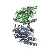
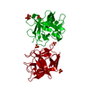
Protein , 1 types, 2 molecules AB

#1: Protein PaiA Acetyltransferase


Mass: 18808.830 Da / Num. of mol.: 2
Source method: isolated from a genetically manipulated source
Source: (gene. exp.) Thermoplasma acidophilum (acidophilic) / Gene: Ta0374 / Plasmid: pMCSG7 / Production host: Escherichia coli BL21 (bacteria) / Strain (production host): Bl21magic / References: UniProt: Q9HL57

-
Non-polymers , 5 types, 82 molecules

#2: Chemical ChemComp-CL / CHLORIDE ION / Chloride


Mass: 35.453 Da / Num. of mol.: 3 / Source method: obtained synthetically / Formula: Cl
#3: Chemical ChemComp-NI / NICKEL (II) ION / Nickel


Mass: 58.693 Da / Num. of mol.: 1 / Source method: obtained synthetically / Formula: Ni
#4: Chemical ChemComp-BR / BROMIDE ION / Bromide


Mass: 79.904 Da / Num. of mol.: 1 / Source method: obtained synthetically / Formula: Br
#5: Chemical ChemComp-ACO / ACETYL COENZYME *A / Acetyl-CoA


Mass: 809.571 Da / Num. of mol.: 2 / Source method: obtained synthetically / Formula: C23H38N7O17P3S
#6: Water ChemComp-HOH / water / Water


Mass: 18.015 Da / Num. of mol.: 75 / Source method: isolated from a natural source / Formula: H2O

-
Experimental details

-
Experiment

ExperimentMethod: X-RAY DIFFRACTION / Number of used crystals: 1

-
Sample preparation

CrystalDensity Matthews: 2.89 Å3/Da / Density % sol: 57.44 %
Crystal growTemperature: 295 K / Method: vapor diffusion, sitting drop / pH: 7.5
Details: 0.2 M Sodium Bromide, 20% PEG 3350, 0.1 M Bis-Tris propane, pH 7.5, VAPOR DIFFUSION, SITTING DROP, temperature 295K

-
Data collection

DiffractionMean temperature: 100 K
Diffraction sourceSource: SYNCHROTRON / Site: APS / Beamline: 21-ID-G / Wavelength: 0.97872 Å
DetectorDetector: CCD / Date: Mar 16, 2009 / Details: MIRROR
RadiationMonochromator: SI-111 CHANNEL / Protocol: MOLECULAR REPLACEMENT / Monochromatic (M) / Laue (L): M / Scattering type: x-ray
Radiation wavelengthWavelength: 0.97872 Å / Relative weight: 1
ReflectionResolution: 2.3→54.98 Å / Num. all: 19708 / Num. obs: 19708 / % possible obs: 98.7 % / Observed criterion σ(I): -3 / Redundancy: 7 % / Rmerge(I) obs: 0.086 / Rsym value: 0.086 / Net I/σ(I): 17.469
Reflection shellResolution: 2.3→2.34 Å / Redundancy: 6.9 % / Rmerge(I) obs: 0.185 / Mean I/σ(I) obs: 10.2 / Rsym value: 0.185 / % possible all: 98.6

-
Processing

Software
NameVersionClassification
Blu-IceMaxdata collection
MOLREPphasing
REFMAC5.5.0051refinement
HKL-2000data reduction
HKL-3000data scaling
RefinementMethod to determine structure: MOLECULAR REPLACEMENT
Starting model: 3F0A
Resolution: 2.3→54.98 Å / Cor.coef. Fo:Fc: 0.944 / Cor.coef. Fo:Fc free: 0.903 / SU B: 10.592 / SU ML: 0.148 / TLS residual ADP flag: LIKELY RESIDUAL / Cross valid method: THROUGHOUT / σ(F): 0 / ESU R: 0.28 / ESU R Free: 0.226 / Stereochemistry target values: MAXIMUM LIKELIHOOD / Details: HYDROGENS HAVE BEEN ADDED IN THE
RfactorNum. reflection% reflectionSelection details
Rfree0.247 1001 5.1 %RANDOM
Rwork0.191 ---
obs0.194 19697 98.57 %-
all-19697 --
Solvent computationIon probe radii: 0.8 Å / Shrinkage radii: 0.8 Å / VDW probe radii: 1.4 Å / Solvent model: BABINET MODEL WITH MASK
Displacement parametersBiso mean: 21.28 Å2
Baniso -1Baniso -2Baniso -3
1--0.73 Å20 Å20 Å2
2--1.25 Å20 Å2
3----0.52 Å2
Refinement stepCycle: LAST / Resolution: 2.3→54.98 Å
ProteinNucleic acidLigandSolventTotal
Num. atoms2652 0 107 75 2834
Refine LS restraints
Refine-IDTypeDev idealDev ideal targetNumber
X-RAY DIFFRACTIONr_bond_refined_d0.0150.0222815
X-RAY DIFFRACTIONr_bond_other_d0.0010.021977
X-RAY DIFFRACTIONr_angle_refined_deg1.4722.0223791
X-RAY DIFFRACTIONr_angle_other_deg0.85834822
X-RAY DIFFRACTIONr_dihedral_angle_1_deg6.0765318
X-RAY DIFFRACTIONr_dihedral_angle_2_deg36.36224.146123
X-RAY DIFFRACTIONr_dihedral_angle_3_deg16.80215556
X-RAY DIFFRACTIONr_dihedral_angle_4_deg22.8851514
X-RAY DIFFRACTIONr_chiral_restr0.0860.2409
X-RAY DIFFRACTIONr_gen_planes_refined0.0050.022963
X-RAY DIFFRACTIONr_gen_planes_other0.0010.02579
X-RAY DIFFRACTIONr_mcbond_it0.9011.51585
X-RAY DIFFRACTIONr_mcbond_other0.2351.5649
X-RAY DIFFRACTIONr_mcangle_it1.74622554
X-RAY DIFFRACTIONr_scbond_it2.85531230
X-RAY DIFFRACTIONr_scangle_it4.5514.51236
Refine LS restraints NCS

Dom-ID: 1 / Ens-ID: 1 / Number: 2278 / Refine-ID: X-RAY DIFFRACTION

Auth asym-IDTypeRms dev position (Å)Weight position
Aloose positional0.75
Bloose thermal2.4310
LS refinement shellResolution: 2.3→2.36 Å / Total num. of bins used: 20
RfactorNum. reflection% reflection
Rfree0.268 78 -
Rwork0.192 1361 -
obs--98.63 %
Refinement TLS params.

Method: refined / Refine-ID: X-RAY DIFFRACTION

IDL112)L122)L132)L222)L232)L332)S11 (Å °)S12 (Å °)S13 (Å °)S21 (Å °)S22 (Å °)S23 (Å °)S31 (Å °)S32 (Å °)S33 (Å °)T112)T122)T132)T222)T232)T332)Origin x (Å)Origin y (Å)Origin z (Å)
12.6662-0.2011-0.51311.71680.14170.91070.01490.03590.1342-0.0506-0.05960.064-0.082-0.12270.04470.01330.01170.00280.0267-0.0250.071312.275-16.789-5.286
21.825-0.1524-0.0762.9756-0.09742.05230.00310.0527-0.117-0.0102-0.0103-0.20150.30360.11310.00730.09180.02370.00920.0285-0.0410.102635.071-40.044-16.584
Refinement TLS group
IDRefine-IDRefine TLS-IDAuth asym-IDAuth seq-ID
1X-RAY DIFFRACTION1A1 - 159
2X-RAY DIFFRACTION2B1 - 159

+
About Yorodumi

-
News

-
Feb 9, 2022. New format data for meta-information of EMDB entries

New format data for meta-information of EMDB entries

  • Version 3 of the EMDB header file is now the official format.
  • The previous official version 1.9 will be removed from the archive.

Related info.:EMDB header

External links:wwPDB to switch to version 3 of the EMDB data model

-
Aug 12, 2020. Covid-19 info

Covid-19 info

URL: https://pdbj.org/emnavi/covid19.php

New page: Covid-19 featured information page in EM Navigator.

Related info.:Covid-19 info / Mar 5, 2020. Novel coronavirus structure data

+
Mar 5, 2020. Novel coronavirus structure data

Novel coronavirus structure data

Related info.:Yorodumi Speices / Aug 12, 2020. Covid-19 info

External links:COVID-19 featured content - PDBj / Molecule of the Month (242):Coronavirus Proteases

+
Jan 31, 2019. EMDB accession codes are about to change! (news from PDBe EMDB page)

EMDB accession codes are about to change! (news from PDBe EMDB page)

  • The allocation of 4 digits for EMDB accession codes will soon come to an end. Whilst these codes will remain in use, new EMDB accession codes will include an additional digit and will expand incrementally as the available range of codes is exhausted. The current 4-digit format prefixed with “EMD-” (i.e. EMD-XXXX) will advance to a 5-digit format (i.e. EMD-XXXXX), and so on. It is currently estimated that the 4-digit codes will be depleted around Spring 2019, at which point the 5-digit format will come into force.
  • The EM Navigator/Yorodumi systems omit the EMD- prefix.

Related info.:Q: What is EMD? / ID/Accession-code notation in Yorodumi/EM Navigator

External links:EMDB Accession Codes are Changing Soon! / Contact to PDBj

+
Jul 12, 2017. Major update of PDB

Major update of PDB

  • wwPDB released updated PDB data conforming to the new PDBx/mmCIF dictionary.
  • This is a major update changing the version number from 4 to 5, and with Remediation, in which all the entries are updated.
  • In this update, many items about electron microscopy experimental information are reorganized (e.g. em_software).
  • Now, EM Navigator and Yorodumi are based on the updated data.

External links:wwPDB Remediation / Enriched Model Files Conforming to OneDep Data Standards Now Available in the PDB FTP Archive

-
Yorodumi

Thousand views of thousand structures

  • Yorodumi is a browser for structure data from EMDB, PDB, SASBDB, etc.
  • This page is also the successor to EM Navigator detail page, and also detail information page/front-end page for Omokage search.
  • The word "yorodu" (or yorozu) is an old Japanese word meaning "ten thousand". "mi" (miru) is to see.

Related info.:EMDB / PDB / SASBDB / Comparison of 3 databanks / Yorodumi Search / Aug 31, 2016. New EM Navigator & Yorodumi / Yorodumi Papers / Jmol/JSmol / Function and homology information / Changes in new EM Navigator and Yorodumi

Read more