 Movie
Movie Controller


+ Open data

- Basic information
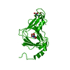
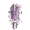
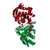
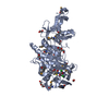
| Entry | Database: PDB / ID: 3k9a | ||||||
|---|---|---|---|---|---|---|---|
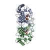
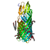
| Title | Crystal Structure of HIV gp41 with MPER | ||||||
 Components Components | HIV glycoprotein gp41 | ||||||
 Keywords Keywords |  VIRAL PROTEIN / HIV / gp41 / membrane proximal external region / MPER VIRAL PROTEIN / HIV / gp41 / membrane proximal external region / MPER | ||||||
| Function / homology | Helix Hairpins - #210 / Helix Hairpins / Orthogonal Bundle / Mainly Alpha Function and homology information Function and homology information | ||||||
| Biological species |   Human immunodeficiency virus 1 Human immunodeficiency virus 1 | ||||||
| Method | X-RAY DIFFRACTION / SYNCHROTRON / Molecular replacement, Br SAD / Resolution: 2.1 Å | ||||||
 Authors Authors | Shi, W. / Han, D. / Habte, H. / Cho, M. / Chance, M.R. | ||||||
 Citation Citation | Journal: J.Biol.Chem. / Year: 2010 Title: Structural characterization of HIV gp41 with the membrane-proximal external region Authors: Shi, W. / Bohon, J. / Han, D.P. / Habte, H. / Qin, Y. / Cho, M.W. / Chance, M.R. | ||||||
| History |
|
- Structure visualization
| Structure viewer | Molecule: MolmilJmol/JSmol |
|---|
- Downloads & links
-Download
| PDBx/mmCIF format | 3k9a.cif.gz | 28.3 KB | Display | PDBx/mmCIF format |
|---|---|---|---|---|
| PDB format | pdb3k9a.ent.gz | 18.3 KB | Display | PDB format |
| PDBx/mmJSON format | 3k9a.json.gz | Tree view | PDBx/mmJSON format | |
| Others |  Other downloads Other downloads |
-Validation report
| Arichive directory | https://data.pdbj.org/pub/pdb/validation_reports/k9/3k9aftp://data.pdbj.org/pub/pdb/validation_reports/k9/3k9a | HTTPS FTP |
|---|
-Related structure data
| Related structure data |  1aikS S: Starting model for refinement |
|---|---|
| Similar structure data |

-Links
 PDBj
PDBj
- Assembly
| Deposited unit | 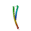
| ||||||||
|---|---|---|---|---|---|---|---|---|---|
| 1 | 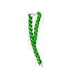
| ||||||||
| Unit cell |
|
-Components
| #1: Protein | Mass: 12722.096 Da / Num. of mol.: 1 / Fragment: gp41 fusion protein / Mutation: HR1+4XGly+HR2+MPER Source method: isolated from a genetically manipulated source Source: (gene. exp.) Human immunodeficiency virus 1 / Strain: MCON6 / Gene: gp41 / Production host:  Escherichia coli (E. coli) Escherichia coli (E. coli) |
|---|---|
| #2: Water | ChemComp-HOH / Water Mass: 18.015 Da / Num. of mol.: 19 / Source method: isolated from a natural source / Formula: H2O Mass: 18.015 Da / Num. of mol.: 19 / Source method: isolated from a natural source / Formula: H2O |
| Sequence details | THIS IS A FUSION PROTEIN WITH A LINKER GGGGS IN THE MIDDLE AND SIX-RESIDUES HIS TAGS AT THE C-TERMINUS END |
-Experimental details
-Experiment
| Experiment | Method: X-RAY DIFFRACTION / Number of used crystals: 1 |
|---|
- Sample preparation
| Crystal | Density Matthews: 2.25 Å3/Da / Density % sol: 45.37 % |
|---|---|
| Crystal grow | Temperature: 298 K / Method: vapor diffusion, sitting drop / pH: 6.5 Details: 45% MPD, 0.2 M sodium acetate, 0.1 M Bis-tris, pH 6.5, VAPOR DIFFUSION, SITTING DROP, temperature 298K |
-Data collection
| Diffraction | Mean temperature: 100 K |
|---|---|
| Diffraction source | Source: SYNCHROTRON / Site: NSLS  / Beamline: X29A / Wavelength: 1.08 Å / Beamline: X29A / Wavelength: 1.08 Å |
| Detector | Type: ADSC QUANTUM 315 / Detector: CCD / Date: Aug 28, 2009 / Details: monochromator and mirror |
| Radiation | Monochromator: SAGITALLY FOCUSED Si(111) / Protocol: SINGLE WAVELENGTH / Monochromatic (M) / Laue (L): M / Scattering type: x-ray |
| Radiation wavelength | Wavelength: 1.08 Å / Relative weight: 1 |
| Reflection | Resolution: 2.1→50 Å / Num. obs: 7537 / % possible obs: 99.8 % / Redundancy: 30 % / Biso Wilson estimate: 40.71 Å2 / Rsym value: 0.074 / Net I/σ(I): 55.8 |
| Reflection shell | Resolution: 2.1→2.14 Å / Redundancy: 29.9 % / Mean I/σ(I) obs: 6 / Num. unique all: 355 / Rsym value: 0.713 / % possible all: 100 |
- Processing
| Software |
| |||||||||||||||||||||||||||||||||||
|---|---|---|---|---|---|---|---|---|---|---|---|---|---|---|---|---|---|---|---|---|---|---|---|---|---|---|---|---|---|---|---|---|---|---|---|---|
| Refinement | Method to determine structure: Molecular replacement, Br SAD Starting model: PDB entry 1AIK Resolution: 2.1→42.2 Å / Isotropic thermal model: isotropic / Cross valid method: THROUGHOUT / σ(F): 1.34
| |||||||||||||||||||||||||||||||||||
| Displacement parameters | Biso mean: 51.4 Å2
| |||||||||||||||||||||||||||||||||||
| Refinement step | Cycle: LAST / Resolution: 2.1→42.2 Å
| |||||||||||||||||||||||||||||||||||
| Refine LS restraints |
| |||||||||||||||||||||||||||||||||||
| LS refinement shell |
|
