| Entry | Database: PDB / ID: 3k8z
|
|---|
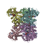
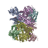
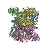
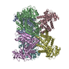
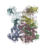
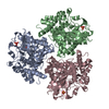
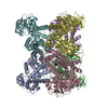
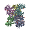
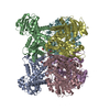
| Title | Crystal Structure of Gudb1 a decryptified secondary glutamate dehydrogenase from B. subtilis |
|---|
 Components Components | NAD-specific glutamate dehydrogenase |
|---|
 Keywords Keywords |  OXIDOREDUCTASE / GudB / Glutamate dehydrogenase / NAD OXIDOREDUCTASE / GudB / Glutamate dehydrogenase / NAD |
|---|
| Function / homology |  Function and homology information Function and homology information
glutamate dehydrogenase / glutamate catabolic process / : / glutamate dehydrogenase (NAD+) activity / amino acid metabolic processSimilarity search - Function Helicase, Ruva Protein; domain 3 - #1210 / Glutamate dehydrogenase / Leucine Dehydrogenase, chain A, domain 1 / NAD(P) binding domain of glutamate dehydrogenase / Leu/Phe/Val dehydrogenases active site / Glu / Leu / Phe / Val dehydrogenases active site. / Glutamate/phenylalanine/leucine/valine dehydrogenase / Glutamate/phenylalanine/leucine/valine dehydrogenase, dimerisation domain / Glu/Leu/Phe/Val dehydrogenase, dimerisation domain / Glutamate/Leucine/Phenylalanine/Valine dehydrogenase ...Helicase, Ruva Protein; domain 3 - #1210 / Glutamate dehydrogenase / Leucine Dehydrogenase, chain A, domain 1 / NAD(P) binding domain of glutamate dehydrogenase / Leu/Phe/Val dehydrogenases active site / Glu / Leu / Phe / Val dehydrogenases active site. / Glutamate/phenylalanine/leucine/valine dehydrogenase / Glutamate/phenylalanine/leucine/valine dehydrogenase, dimerisation domain / Glu/Leu/Phe/Val dehydrogenase, dimerisation domain / Glutamate/Leucine/Phenylalanine/Valine dehydrogenase / Glutamate/phenylalanine/leucine/valine dehydrogenase, C-terminal / Glutamate/Leucine/Phenylalanine/Valine dehydrogenase / Helicase, Ruva Protein; domain 3 / NAD(P)-binding Rossmann-like Domain / NAD(P)-binding domain superfamily / Rossmann fold / Orthogonal Bundle / 3-Layer(aba) Sandwich / Mainly Alpha / Alpha BetaSimilarity search - Domain/homology |
|---|
| Biological species |   Bacillus subtilis (bacteria) Bacillus subtilis (bacteria) |
|---|
| Method | X-RAY DIFFRACTION / SYNCHROTRON / MOLECULAR REPLACEMENT / molecular replacement / Resolution: 2.4 Å |
|---|
 Authors Authors | Gunka, K. / Newman, J.A. / Commichau, F.M. / Herzberg, C. / Rodrigues, C. / Hewitt, L. / Lewis, R.J. / Stulke, J. |
|---|
 Citation Citation | Journal: J.Mol.Biol. / Year: 2010
Title: Functional dissection of a trigger enzyme: mutations of the bacillus subtilis glutamate dehydrogenase RocG that affect differentially its catalytic activity and regulatory properties
Authors: Gunka, K. / Newman, J.A. / Commichau, F.M. / Herzberg, C. / Rodrigues, C. / Hewitt, L. / Lewis, R.J. / Stulke, J. |
|---|
| History | | Deposition | Oct 15, 2009 | Deposition site: RCSB / Processing site: PDBJ |
|---|
| Revision 1.0 | Jun 2, 2010 | Provider: repository / Type: Initial release |
|---|
| Revision 1.1 | Jul 13, 2011 | Group: Version format compliance |
|---|
| Revision 1.2 | Jan 22, 2014 | Group: Database references |
|---|
| Revision 1.3 | Mar 20, 2024 | Group: Data collection / Database references / Refinement description
Category: chem_comp_atom / chem_comp_bond ...chem_comp_atom / chem_comp_bond / database_2 / struct_ncs_dom_lim
Item: _database_2.pdbx_DOI / _database_2.pdbx_database_accession ..._database_2.pdbx_DOI / _database_2.pdbx_database_accession / _struct_ncs_dom_lim.beg_auth_comp_id / _struct_ncs_dom_lim.beg_label_asym_id / _struct_ncs_dom_lim.beg_label_comp_id / _struct_ncs_dom_lim.beg_label_seq_id / _struct_ncs_dom_lim.end_auth_comp_id / _struct_ncs_dom_lim.end_label_asym_id / _struct_ncs_dom_lim.end_label_comp_id / _struct_ncs_dom_lim.end_label_seq_id |
|---|
|
|---|
 Movie
Movie Controller

 Yorodumi
 Open data
 Basic information
 Structure visualization
 Downloads & links
 Other downloads

 PDBj
PDBj Assembly