 Movie
Movie Controller

[English] 日本語
 Yorodumi
- PDB-3k5h: Crystal structure of carboxyaminoimidazole ribonucleotide synthas... -
+ Open data

- Basic information
| Entry | Database: PDB / ID: 3k5h | ||||||
|---|---|---|---|---|---|---|---|
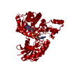
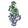
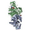
| Title | Crystal structure of carboxyaminoimidazole ribonucleotide synthase from asperigillus clavatus complexed with ATP | ||||||
 Components Components | Phosphoribosyl-aminoimidazole carboxylase | ||||||
 Keywords Keywords |  LYASE / purine biosynthesis / ATP-grasp LYASE / purine biosynthesis / ATP-grasp | ||||||
| Function / homology |  Function and homology informationphosphoribosylaminoimidazole carboxylase / 5-amino-4-imidazole carboxylate lyase activity / phosphoribosylaminoimidazole carboxylase activity / 'de novo' IMP biosynthetic process / ATP binding / metal ion binding Function and homology informationphosphoribosylaminoimidazole carboxylase / 5-amino-4-imidazole carboxylate lyase activity / phosphoribosylaminoimidazole carboxylase activity / 'de novo' IMP biosynthetic process / ATP binding / metal ion bindingSimilarity search - Function | ||||||
| Biological species |  Aspergillus clavatus (mold) Aspergillus clavatus (mold) | ||||||
| Method | X-RAY DIFFRACTION / MOLECULAR REPLACEMENT / Resolution: 2.1 Å | ||||||
 Authors Authors | Thoden, J.B. / Holden, H.M. / Paritala, H. / Firestine, S.M. | ||||||
 Citation Citation | Journal: Biochemistry / Year: 2010 Title: Structural and functional studies of Aspergillus clavatus N(5)-carboxyaminoimidazole ribonucleotide synthetase Authors: Thoden, J.B. / Holden, H.M. / Paritala, H. / Firestine, S.M. | ||||||
| History |
|
- Structure visualization
| Structure viewer | Molecule: MolmilJmol/JSmol |
|---|
- Downloads & links
-Download
| PDBx/mmCIF format | 3k5h.cif.gz | 329.2 KB | Display | PDBx/mmCIF format |
|---|---|---|---|---|
| PDB format | pdb3k5h.ent.gz | 264 KB | Display | PDB format |
| PDBx/mmJSON format | 3k5h.json.gz | Tree view | PDBx/mmJSON format | |
| Others |  Other downloads Other downloads |
-Validation report
| Arichive directory | https://data.pdbj.org/pub/pdb/validation_reports/k5/3k5hftp://data.pdbj.org/pub/pdb/validation_reports/k5/3k5h | HTTPS FTP |
|---|
-Related structure data


-Links
 PDBj
PDBj


- Assembly
| Deposited unit | 
| ||||||||
|---|---|---|---|---|---|---|---|---|---|
| 1 | 
| ||||||||
| 2 | 
| ||||||||
| Unit cell |
|
-Components
| #1: Protein | Mass: 44720.836 Da / Num. of mol.: 4 / Fragment: residues 1-383 Source method: isolated from a genetically manipulated source Source: (gene. exp.) Aspergillus clavatus (mold) / Strain: NRRL 1 / Gene: ACLA_051590, ade2 / Plasmid: pET28 / Production host:  Escherichia coli (E. coli) / Strain (production host): Rosettas(DE2) Escherichia coli (E. coli) / Strain (production host): Rosettas(DE2)References: UniProt: A1CII2, phosphoribosylaminoimidazole carboxylase#2: Chemical | ChemComp-ATP / Adenosine triphosphate  Mass: 507.181 Da / Num. of mol.: 4 / Source method: obtained synthetically / Formula: C10H16N5O13P3 / Comment: ATP, energy-carrying molecule*YM Mass: 507.181 Da / Num. of mol.: 4 / Source method: obtained synthetically / Formula: C10H16N5O13P3 / Comment: ATP, energy-carrying molecule*YM#3: Chemical | ChemComp-MG /   Mass: 24.305 Da / Num. of mol.: 8 / Source method: obtained synthetically / Formula: Mg Mass: 24.305 Da / Num. of mol.: 8 / Source method: obtained synthetically / Formula: Mg#4: Water | ChemComp-HOH / | Water Mass: 18.015 Da / Num. of mol.: 849 / Source method: isolated from a natural source / Formula: H2O Mass: 18.015 Da / Num. of mol.: 849 / Source method: isolated from a natural source / Formula: H2O |
|---|
-Experimental details
-Experiment
| Experiment | Method: X-RAY DIFFRACTION / Number of used crystals: 1 |
|---|
- Sample preparation
| Crystal | Density Matthews: 2.72 Å3/Da / Density % sol: 54.71 % |
|---|---|
| Crystal grow | Temperature: 298 K / Method: vapor diffusion, hanging drop / pH: 9 Details: 18% PEG-5000 monomethylether, 10 mM ATP, 40 mM MgCl2, 200 mM NaCl, 100mM CHES, pH 9, VAPOR DIFFUSION, HANGING DROP, temperature 298K |
-Data collection
| Diffraction | Mean temperature: 100 K |
|---|---|
| Diffraction source | Source: ROTATING ANODE / Type: RIGAKU RU200 / Wavelength: 1.5418 Å |
| Detector | Type: Bruker Platinum 135 / Detector: CCD / Date: Jan 14, 2009 / Details: montel |
| Radiation | Monochromator: Ni filter / Protocol: SINGLE WAVELENGTH / Monochromatic (M) / Laue (L): M / Scattering type: x-ray |
| Radiation wavelength | Wavelength: 1.5418 Å / Relative weight: 1 |
| Reflection | Resolution: 2.1→30 Å / Num. all: 104348 / Num. obs: 104348 / % possible obs: 93.6 % / Observed criterion σ(F): 0 / Observed criterion σ(I): 0 / Redundancy: 4 % / Rmerge(I) obs: 0.087 / Rsym value: 0.087 / Net I/σ(I): 9.6 |
| Reflection shell | Resolution: 2.1→2.2 Å / Redundancy: 1.8 % / Rmerge(I) obs: 0.309 / Mean I/σ(I) obs: 2.2 / Num. unique all: 12740 / Rsym value: 0.309 / % possible all: 86.5 |
- Processing
| Software |
| |||||||||||||||||||||||||
|---|---|---|---|---|---|---|---|---|---|---|---|---|---|---|---|---|---|---|---|---|---|---|---|---|---|---|
| Refinement | Method to determine structure: MOLECULAR REPLACEMENT Starting model: non-published structure (in-house) from yeast Resolution: 2.1→30 Å / Isotropic thermal model: isotropic / Cross valid method: THROUGHOUT / σ(F): 0 / σ(I): 0 / Stereochemistry target values: Engh & Huber
| |||||||||||||||||||||||||
| Refinement step | Cycle: LAST / Resolution: 2.1→30 Å
|
