 Movie
Movie Controller

[English] 日本語
 Yorodumi
- PDB-3k20: X-ray structure of oxidoreductase from corynebacterium diphtheria... -
+ Open data

- Basic information
| Entry | Database: PDB / ID: 3k20 | ||||||
|---|---|---|---|---|---|---|---|
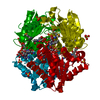
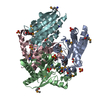
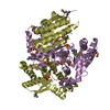
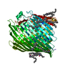
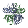
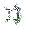
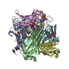
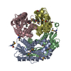
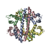
| Title | X-ray structure of oxidoreductase from corynebacterium diphtheriae,hexagonal crystal form. northeast structural genomics consortium target cdr100d | ||||||
 Components Components | oxidoreductase | ||||||
 Keywords Keywords | OXIDOREDUCTASE / Structural Genomics / PSI-2 / Protein Structure Initiative / Northeast Structural Genomics Consortium / NESG / CdR100D / Q6NGSI_CORDI | ||||||
| Function / homology |  Function and homology information Function and homology information | ||||||
| Biological species |  Corynebacterium diphtheriae (bacteria) Corynebacterium diphtheriae (bacteria) | ||||||
| Method | X-RAY DIFFRACTION / SYNCHROTRON / MAD / Resolution: 2.5 Å | ||||||
 Authors Authors | Kuzin, A. / Lew, S. / Sahdev, S. / Xiao, R. / Ciccosanti, C. / Wang, H. / Everett, J.K. / Nair, R. / Acton, T.B. / Rost, B. ...Kuzin, A. / Lew, S. / Sahdev, S. / Xiao, R. / Ciccosanti, C. / Wang, H. / Everett, J.K. / Nair, R. / Acton, T.B. / Rost, B. / Montelione, G.T. / Tong, L. / Hunt, J.F. / Northeast Structural Genomics Consortium (NESG) | ||||||
 Citation Citation | Journal: To be Published Title: Northeast Structural Genomics Consortium Target CdR100D Authors: Kuzin, A. / Lew, S. / Seetharaman, J. / Sahdev, S. / Xiao, R. / Ciccosanti, C. / Wang, H. / Everett, J.K. / Nair, R. / Acton, T.B. / Rost, B. / Montelione, G.T. / Tong, L. / Hunt, J.F. | ||||||
| History |
|
- Structure visualization
| Structure viewer | Molecule: MolmilJmol/JSmol |
|---|
- Downloads & links
-Download
| PDBx/mmCIF format | 3k20.cif.gz | 78.4 KB | Display | PDBx/mmCIF format |
|---|---|---|---|---|
| PDB format | pdb3k20.ent.gz | 63.4 KB | Display | PDB format |
| PDBx/mmJSON format | 3k20.json.gz | Tree view | PDBx/mmJSON format | |
| Others |  Other downloads Other downloads |
-Validation report
| Arichive directory | https://data.pdbj.org/pub/pdb/validation_reports/k2/3k20ftp://data.pdbj.org/pub/pdb/validation_reports/k2/3k20 | HTTPS FTP |
|---|
-Related structure data

-Links
 PDBj
PDBj- Assembly
| Deposited unit | 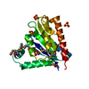
| ||||||||
|---|---|---|---|---|---|---|---|---|---|
| 1 | 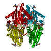
| ||||||||
| Unit cell |
| ||||||||
| Components on special symmetry positions |
|
-Components
| #1: Protein | Mass: 20457.164 Da / Num. of mol.: 1 Source method: isolated from a genetically manipulated source Source: (gene. exp.) Corynebacterium diphtheriae (bacteria) / Gene: DIP1437 / Plasmid: pET 14-15C / Production host: Escherichia coli BL21(DE3) (bacteria) / Strain (production host): BL21(DE3)+ Magic / References: UniProt: Q6NGS1 | ||
|---|---|---|---|
| #2: Chemical | ChemComp-SO4 / Sulfate  Mass: 96.063 Da / Num. of mol.: 4 / Source method: obtained synthetically / Formula: SO4 Mass: 96.063 Da / Num. of mol.: 4 / Source method: obtained synthetically / Formula: SO4#3: Water | ChemComp-HOH / | Water Mass: 18.015 Da / Num. of mol.: 33 / Source method: isolated from a natural source / Formula: H2O Mass: 18.015 Da / Num. of mol.: 33 / Source method: isolated from a natural source / Formula: H2O |
-Experimental details
-Experiment
| Experiment | Method: X-RAY DIFFRACTION / Number of used crystals: 1 |
|---|
- Sample preparation
| Crystal | Density Matthews: 4.83 Å3/Da / Density % sol: 74.53 % |
|---|---|
| Crystal grow | pH: 7.5 Details: Protein solution: 100mM NaCl, 5mM DTT, 0.02% NaN3, 10mM Tris-HCl (pH 7.5) . Reservoir solution:2.2M ammonium sulfate, 0.1M sodium acetate, 3% ethanol, VAPOR DIFFUSION, HANGING DROP |
-Data collection
| Diffraction | Mean temperature: 100 K | ||||||||||||
|---|---|---|---|---|---|---|---|---|---|---|---|---|---|
| Diffraction source | Source: SYNCHROTRON / Site: SSRL  / Beamline: BL9-2 / Wavelength: 0.97879 0.91837 0.97926 / Beamline: BL9-2 / Wavelength: 0.97879 0.91837 0.97926 | ||||||||||||
| Detector | Type: MARMOSAIC 325 mm CCD / Detector: CCD / Date: Aug 4, 2009 Details: Si(111). A SECOND SET OF Si(220) crystals is also available. | ||||||||||||
| Radiation | Monochromator: DOUBLE CRYSTAL MONOCHROMATOR / Protocol: MAD / Monochromatic (M) / Laue (L): M / Scattering type: x-ray | ||||||||||||
| Radiation wavelength |
| ||||||||||||
| Reflection | Resolution: 2.5→30 Å / Num. obs: 26206 / % possible obs: 98.7 % / Observed criterion σ(I): -3 / Redundancy: 3.8 % / Rmerge(I) obs: 0.066 / Net I/σ(I): 19.1 | ||||||||||||
| Reflection shell | Resolution: 2.5→2.59 Å / Redundancy: 3.4 % / Rmerge(I) obs: 0.311 / Mean I/σ(I) obs: 2.9 / % possible all: 98.7 |
- Processing
| Software |
| ||||||||||||||||||||||||||||||||||||||||||
|---|---|---|---|---|---|---|---|---|---|---|---|---|---|---|---|---|---|---|---|---|---|---|---|---|---|---|---|---|---|---|---|---|---|---|---|---|---|---|---|---|---|---|---|
| Refinement | Method to determine structure: MAD / Resolution: 2.5→29.51 Å / SU ML: 0.35 / Phase error: 25.13 / Stereochemistry target values: ML
| ||||||||||||||||||||||||||||||||||||||||||
| Solvent computation | Shrinkage radii: 0.9 Å / Solvent model: FLAT BULK SOLVENT MODEL | ||||||||||||||||||||||||||||||||||||||||||
| Displacement parameters | Biso mean: 50.06 Å2 | ||||||||||||||||||||||||||||||||||||||||||
| Refinement step | Cycle: LAST / Resolution: 2.5→29.51 Å
| ||||||||||||||||||||||||||||||||||||||||||
| Refine LS restraints |
| ||||||||||||||||||||||||||||||||||||||||||
| LS refinement shell |
| ||||||||||||||||||||||||||||||||||||||||||
| Refinement TLS params. | Method: refined / Origin x: 33.3447 Å / Origin y: 31.5544 Å / Origin z: 35.8693 Å
| ||||||||||||||||||||||||||||||||||||||||||
| Refinement TLS group | Selection details: all |
