 Movie
Movie Controller

[English] 日本語
 Yorodumi
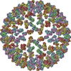
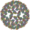
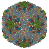
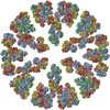
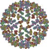
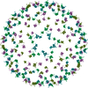
- PDB-3k1q: Backbone model of an aquareovirus virion by cryo-electron microsc... -
+ Open data

- Basic information
| Entry | Database: PDB / ID: 3k1q | ||||||
|---|---|---|---|---|---|---|---|
| Title | Backbone model of an aquareovirus virion by cryo-electron microscopy and bioinformatics | ||||||
 Components Components |
| ||||||
 Keywords Keywords |  VIRUS / cryoEM / bioinformatics / structure-based refinement / macromolecular assembly / GCRV virion VIRUS / cryoEM / bioinformatics / structure-based refinement / macromolecular assembly / GCRV virion | ||||||
| Function / homology |  Function and homology information Function and homology informationhost cell surface binding / viral inner capsid / viral outer capsid / permeabilization of host organelle membrane involved in viral entry into host cell / symbiont entry into host cell via permeabilization of inner membrane / 7-methylguanosine mRNA capping / viral capsid / mRNA guanylyltransferase activity / mRNA 5'-cap (guanine-N7-)-methyltransferase activity / RNA helicase activity ...host cell surface binding / viral inner capsid / viral outer capsid / permeabilization of host organelle membrane involved in viral entry into host cell / symbiont entry into host cell via permeabilization of inner membrane / 7-methylguanosine mRNA capping / viral capsid / mRNA guanylyltransferase activity / mRNA 5'-cap (guanine-N7-)-methyltransferase activity / RNA helicase activity / RNA helicase / hydrolase activity / GTP binding / ATP binding / metal ion bindingSimilarity search - Function | ||||||
| Biological species |  Grass carp reovirus Grass carp reovirus | ||||||
| Method | ELECTRON MICROSCOPY / single particle reconstruction / cryo EM / Resolution: 4.5 Å | ||||||
 Authors Authors | Cheng, L.P. / Zhu, J. / Hiu, W.H. / Zhang, X.K. / Honig, B. / Fang, Q. / Zhou, Z.H. | ||||||
 Citation Citation | Journal: J Mol Biol / Year: 2010 Title: Backbone model of an aquareovirus virion by cryo-electron microscopy and bioinformatics. Authors: Lingpeng Cheng / Jiang Zhu / Wong Hoi Hui / Xiaokang Zhang / Barry Honig / Qin Fang / Z Hong Zhou /  Abstract: Grass carp reovirus (GCRV) is a member of the aquareovirus genus in the Reoviridae family and has a capsid with two shells-a transcription-competent core surrounded by a coat. We report a near-atomic- ...Grass carp reovirus (GCRV) is a member of the aquareovirus genus in the Reoviridae family and has a capsid with two shells-a transcription-competent core surrounded by a coat. We report a near-atomic-resolution reconstruction of the GCRV virion by cryo-electron microscopy and single-particle reconstruction. A backbone model of the GCRV virion, including seven conformers of the five capsid proteins making up the 1500 molecules in both the core and the coat, was derived using cryo-electron microscopy density-map-constrained homology modeling and refinement. Our structure clearly showed that the amino-terminal segment of core protein VP3B forms an approximately 120-A-long alpha-helix-rich extension bridging across the icosahedral 2-fold-symmetry-related molecular interface. The presence of this unique structure across this interface and the lack of an external cementing molecule at this location in GCRV suggest a stabilizing role of this extended amino-terminal density. Moreover, part of this amino-terminal extension becomes invisible in the reconstruction of transcription-competent core particles, suggesting its involvement in endogenous viral RNA transcription. Our structure of the VP1 turret represents its open state, and comparison with its related structures at the closed state suggests hinge-like domain movements associated with the mRNA-capping machinery. Overall, this first backbone model of an aquareovirus virion provides a wealth of structural information for understanding the structural basis of GCRV assembly and transcription. #1: Journal: To be PublishedTitle: Building Models for Molecular Assemblies by Cryo-electron Microscopy, Homology Modeling and Multi-scale Structure-Based Refinement Authors: Honig, B. | ||||||
| History |
|
- Structure visualization
| Movie |
Movie viewer |
|---|---|
| Structure viewer | Molecule: MolmilJmol/JSmol |
- Downloads & links
-Download
| PDBx/mmCIF format | 3k1q.cif.gz | 2 MB | Display | PDBx/mmCIF format |
|---|---|---|---|---|
| PDB format | pdb3k1q.ent.gz | Display | PDB format | |
| PDBx/mmJSON format | 3k1q.json.gz | Tree view | PDBx/mmJSON format | |
| Others |  Other downloads Other downloads |
-Validation report
| Arichive directory | https://data.pdbj.org/pub/pdb/validation_reports/k1/3k1qftp://data.pdbj.org/pub/pdb/validation_reports/k1/3k1q | HTTPS FTP |
|---|
-Related structure data
| Related structure data | 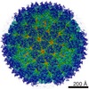 1653MC M: map data used to model this data C: citing same article ( |
|---|---|
| Similar structure data |


-Links
 PDBj
PDBj


- Assembly
| Deposited unit | 
|
|---|---|
| 1 | x 60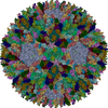
|
| 2 |
|
| 3 | x 5
|
| 4 | x 6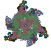
|
| 5 | 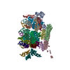
|
| Symmetry | Point symmetry: (Schoenflies symbol: I (icosahedral)) |
-Components
-Protein , 4 types, 5 molecules ABCDE
| #1: Protein | Mass: 141512.156 Da / Num. of mol.: 1 / Source method: isolated from a natural source / Source: (natural) Grass carp reovirus / References: UniProt: Q9E3W0 |
|---|---|
| #2: Protein | Mass: 112883.250 Da / Num. of mol.: 1 / Source method: isolated from a natural source / Source: (natural) Grass carp reovirus / References: UniProt: Q9E3V8 |
| #3: Protein | Mass: 130304.164 Da / Num. of mol.: 1 / Source method: isolated from a natural source / Source: (natural) Grass carp reovirus / References: UniProt: Q9E3V8 |
| #4: Protein | Mass: 44606.535 Da / Num. of mol.: 2 / Source method: isolated from a natural source / Source: (natural) Grass carp reovirus / References: UniProt: Q8JU64 |
-Outer capsid ... , 2 types, 20 molecules FGHLMNRSTYIJKOPQUVWX
| #5: Protein | Mass: 29844.648 Da / Num. of mol.: 10 / Source method: isolated from a natural source / Source: (natural) Grass carp reovirus / References: UniProt: Q8JU63#6: Protein | Mass: 67715.734 Da / Num. of mol.: 10 / Source method: isolated from a natural source / Source: (natural) Grass carp reovirus / References: UniProt: Q8JU67 |
|---|
-Experimental details
-Experiment
| Experiment | Method: ELECTRON MICROSCOPY |
|---|---|
| EM experiment | Aggregation state: PARTICLE / 3D reconstruction method: single particle reconstruction |
- Sample preparation
| Component | Name: Aqaurevirus: Grass carp reovirus (GCRV) / Type: VIRUS / Synonym: sputnik |
|---|---|
| Details of virus | Host category: CARP / Type: VIRION |
| Natural host | Organism: Ctenopharyngodon idella |
| Buffer solution | Details: 10mM phosphate-buffered saline |
| Specimen | Conc.: 1 mg/ml / Embedding applied: NO / Shadowing applied: NO / Staining applied: NO / Vitrification applied: YES |
| Specimen support | Details: Quantifoil holey carbon grids, 2um/1um type |
| Vitrification | Cryogen name: ETHANE / Details: Manual pluger freshing into liquid ethane |
- Electron microscopy imaging
| Experimental equipment |  Model: Tecnai Polara / Image courtesy: FEI Company |
|---|---|
| Microscopy | Model: FEI POLARA 300 Details: The electron beam is carefully monitored to ensure that the beam is just slightly larger than the CCD. See above |
| Electron gun | Electron source: FIELD EMISSION GUN / Accelerating voltage: 300 kV / Illumination mode: FLOOD BEAM |
| Electron lens | Mode: BRIGHT FIELDBright-field microscopy / Nominal magnification: 154380 X / Nominal defocus max: 2300 nm / Nominal defocus min: 800 nm / Cs: 2 mm |
| Specimen holder | Temperature: 77 K / Tilt angle max: 0 ° / Tilt angle min: 0 ° |
| Image recording | Electron dose: 20 e/Å2 / Film or detector model: GENERIC CCD |
| Image scans | Num. digital images: 4000 |
| Radiation | Protocol: SINGLE WAVELENGTH / Monochromatic (M) / Laue (L): M / Scattering type: x-ray |
| Radiation wavelength | Relative weight: 1 |
- Processing
| CTF correction | Details: Fully corrected. See Zhou et al., 1999, J. Virol. 73, 3210-3218 | ||||||||||||
|---|---|---|---|---|---|---|---|---|---|---|---|---|---|
| Symmetry | Point symmetry: I (icosahedral) | ||||||||||||
| 3D reconstruction | Resolution: 4.5 Å / Num. of particles: 15000 / Nominal pixel size: 0.97 Å / Actual pixel size: 0.97 Å / Symmetry type: POINT | ||||||||||||
| Atomic model building | B value: 100 | ||||||||||||
| Refinement step | Cycle: LAST
|
