 Movie
Movie Controller

[English] 日本語
 Yorodumi
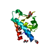
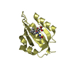
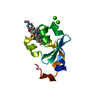
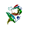
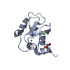
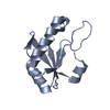
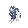
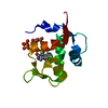
- PDB-3k0x: Crystal structure of telomere capping protein Ten1 from Saccharom... -
+ Open data

- Basic information
| Entry | Database: PDB / ID: 3k0x | ||||||
|---|---|---|---|---|---|---|---|
| Title | Crystal structure of telomere capping protein Ten1 from Saccharomyces pombe | ||||||
 Components Components | Protein Ten1 | ||||||
 Keywords Keywords |  PROTEIN BINDING / beta barrel / ob fold / telomere capping / Chromosomal protein / Nucleus / Telomere PROTEIN BINDING / beta barrel / ob fold / telomere capping / Chromosomal protein / Nucleus / Telomere | ||||||
| Function / homology |  Function and homology informationCST complex / telomere cap complex / chromosome, telomeric repeat region / single-stranded telomeric DNA binding / telomere capping Function and homology informationCST complex / telomere cap complex / chromosome, telomeric repeat region / single-stranded telomeric DNA binding / telomere cappingSimilarity search - Function | ||||||
| Biological species |  Schizosaccharomyces pombe (fission yeast) Schizosaccharomyces pombe (fission yeast) | ||||||
| Method | X-RAY DIFFRACTION / SAD / Resolution: 1.7 Å | ||||||
 Authors Authors | Gelinas, A.D. / Reyes, F.E. / Batey, R.T. / Wuttke, D.S. | ||||||
 Citation Citation | Journal: Proc.Natl.Acad.Sci.USA / Year: 2009 Title: Telomere capping proteins are structurally related to RPA with an additional telomere-specific domain. Authors: Gelinas, A.D. / Paschini, M. / Reyes, F.E. / Heroux, A. / Batey, R.T. / Lundblad, V. / Wuttke, D.S. | ||||||
| History |
|
- Structure visualization
| Structure viewer | Molecule: MolmilJmol/JSmol |
|---|
- Downloads & links
-Download
| PDBx/mmCIF format | 3k0x.cif.gz | 36.3 KB | Display | PDBx/mmCIF format |
|---|---|---|---|---|
| PDB format | pdb3k0x.ent.gz | 23.8 KB | Display | PDB format |
| PDBx/mmJSON format | 3k0x.json.gz | Tree view | PDBx/mmJSON format | |
| Others |  Other downloads Other downloads |
-Validation report
| Arichive directory | https://data.pdbj.org/pub/pdb/validation_reports/k0/3k0xftp://data.pdbj.org/pub/pdb/validation_reports/k0/3k0x | HTTPS FTP |
|---|
-Related structure data


-Links
 PDBj
PDBj
- Assembly
| Deposited unit | 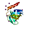
| ||||||||
|---|---|---|---|---|---|---|---|---|---|
| 1 |
| ||||||||
| Unit cell |
| ||||||||
| Components on special symmetry positions |
|
-Components
| #1: Protein | Mass: 11549.443 Da / Num. of mol.: 1 Source method: isolated from a genetically manipulated source Source: (gene. exp.) Schizosaccharomyces pombe (fission yeast)Gene: SPCC1393.14, ten1 / Plasmid: pET duet / Production host:  Escherichia coli (E. coli) / Strain (production host): BL21(DE3) / References: UniProt: P0C5Y7 Escherichia coli (E. coli) / Strain (production host): BL21(DE3) / References: UniProt: P0C5Y7 | ||
|---|---|---|---|
| #2: Chemical | ChemComp-IOD / Iodide  Mass: 126.904 Da / Num. of mol.: 8 / Source method: obtained synthetically / Formula: I Mass: 126.904 Da / Num. of mol.: 8 / Source method: obtained synthetically / Formula: I#3: Water | ChemComp-HOH / | Water Mass: 18.015 Da / Num. of mol.: 146 / Source method: isolated from a natural source / Formula: H2O Mass: 18.015 Da / Num. of mol.: 146 / Source method: isolated from a natural source / Formula: H2O |
-Experimental details
-Experiment
| Experiment | Method: X-RAY DIFFRACTION / Number of used crystals: 1 |
|---|
- Sample preparation
| Crystal | Density Matthews: 2.28 Å3/Da / Density % sol: 46.1 % |
|---|---|
| Crystal grow | Temperature: 290 K / Method: vapor diffusion, hanging drop / pH: 7.8 Details: 0.2 M Potassium nitrate, 25% PEG 3350, pH 7.8, VAPOR DIFFUSION, HANGING DROP, temperature 290K |
-Data collection
| Diffraction | Mean temperature: 93.15 K | ||||||||||||||||||||||||||||||||||||||||||||||||||||||||||||||||||||||||||||||||||||||||
|---|---|---|---|---|---|---|---|---|---|---|---|---|---|---|---|---|---|---|---|---|---|---|---|---|---|---|---|---|---|---|---|---|---|---|---|---|---|---|---|---|---|---|---|---|---|---|---|---|---|---|---|---|---|---|---|---|---|---|---|---|---|---|---|---|---|---|---|---|---|---|---|---|---|---|---|---|---|---|---|---|---|---|---|---|---|---|---|---|---|
| Diffraction source | Source: ROTATING ANODE / Type: RIGAKU / Wavelength: 1.502 Å | ||||||||||||||||||||||||||||||||||||||||||||||||||||||||||||||||||||||||||||||||||||||||
| Detector | Type: RIGAKU RAXIS IV / Detector: IMAGE PLATE / Date: May 8, 2009 | ||||||||||||||||||||||||||||||||||||||||||||||||||||||||||||||||||||||||||||||||||||||||
| Radiation | Monochromator: Ni FILTER / Protocol: SINGLE WAVELENGTH / Monochromatic (M) / Laue (L): M / Scattering type: x-ray | ||||||||||||||||||||||||||||||||||||||||||||||||||||||||||||||||||||||||||||||||||||||||
| Radiation wavelength | Wavelength: 1.502 Å / Relative weight: 1 | ||||||||||||||||||||||||||||||||||||||||||||||||||||||||||||||||||||||||||||||||||||||||
| Reflection | Resolution: 1.7→22.08 Å / Num. obs: 10634 / % possible obs: 94.5 % / Redundancy: 4.5 % / Rsym value: 0.048 / Net I/σ(I): 10.205 | ||||||||||||||||||||||||||||||||||||||||||||||||||||||||||||||||||||||||||||||||||||||||
| Reflection shell |
|
-Phasing
| Phasing | Method: SAD | |||||||||||||||||||||||||||||||||||
|---|---|---|---|---|---|---|---|---|---|---|---|---|---|---|---|---|---|---|---|---|---|---|---|---|---|---|---|---|---|---|---|---|---|---|---|---|
| Phasing MAD | D res high: 1.59 Å / D res low: 25.67 Å / FOM : 0.568 / FOM acentric: 0.558 / FOM centric: 0 / Reflection: 11363 / Reflection acentric: 10395 / Reflection centric: 0 | |||||||||||||||||||||||||||||||||||
| Phasing dm | FOM : 0.74 / FOM acentric: 0.74 / FOM centric: 0 / Reflection: 11366 / Reflection acentric: 11366 / Reflection centric: 0 | |||||||||||||||||||||||||||||||||||
| Phasing dm shell |
|
- Processing
| Software |
| ||||||||||||||||||||||||||||||||
|---|---|---|---|---|---|---|---|---|---|---|---|---|---|---|---|---|---|---|---|---|---|---|---|---|---|---|---|---|---|---|---|---|---|
| Refinement | Method to determine structure: SAD / Resolution: 1.7→20 Å / Occupancy max: 1 / Occupancy min: 0.3 / σ(F): 0
| ||||||||||||||||||||||||||||||||
| Solvent computation | Bsol: 66.185 Å2 | ||||||||||||||||||||||||||||||||
| Displacement parameters | Biso max: 77.25 Å2 / Biso mean: 24.188 Å2 / Biso min: 2.81 Å2
| ||||||||||||||||||||||||||||||||
| Refine analyze | Luzzati coordinate error obs: 0.21 Å / Luzzati d res low obs: 5 Å / Luzzati sigma a obs: 0.27 Å | ||||||||||||||||||||||||||||||||
| Refinement step | Cycle: LAST / Resolution: 1.7→20 Å
| ||||||||||||||||||||||||||||||||
| Refine LS restraints |
| ||||||||||||||||||||||||||||||||
| LS refinement shell | Resolution: 1.7→1.73 Å
| ||||||||||||||||||||||||||||||||
| Xplor file |
|
