 Movie
Movie Controller


+ Open data

- Basic information
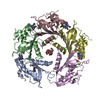
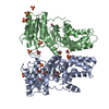
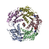
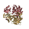
| Entry | Database: PDB / ID: 3jv1 | ||||||
|---|---|---|---|---|---|---|---|
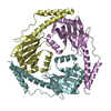
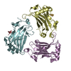
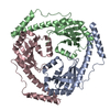
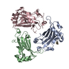
| Title | Crystal structure of the Trypanosoma brucei p22 protein | ||||||
 Components Components | P22 protein | ||||||
 Keywords Keywords |  HYDROLASE / Mam33 family HYDROLASE / Mam33 family | ||||||
| Function / homology |  Function and homology informationRNA modification / translation activator activity / mitochondrial ribosome binding / positive regulation of mitochondrial translation / mitochondrial matrix / mRNA binding / mitochondrion Function and homology informationRNA modification / translation activator activity / mitochondrial ribosome binding / positive regulation of mitochondrial translation / mitochondrial matrix / mRNA binding / mitochondrionSimilarity search - Function | ||||||
| Biological species |  Trypanosoma brucei (eukaryote) Trypanosoma brucei (eukaryote) | ||||||
| Method | X-RAY DIFFRACTION / MOLECULAR REPLACEMENT / Resolution: 2 Å | ||||||
 Authors Authors | Sprehe, M. / Read, L.K. / Schumacher, M.A. | ||||||
 Citation Citation | Journal: J.Biol.Chem. / Year: 2010 Title: Structure of the Trypanosoma brucei p22 protein, a cytochrome oxidase subunit II-specific RNA-editing accessory factor. Authors: Sprehe, M. / Fisk, J.C. / McEvoy, S.M. / Read, L.K. / Schumacher, M.A. | ||||||
| History |
|
- Structure visualization
| Structure viewer | Molecule: MolmilJmol/JSmol |
|---|
- Downloads & links
-Download
| PDBx/mmCIF format | 3jv1.cif.gz | 48.1 KB | Display | PDBx/mmCIF format |
|---|---|---|---|---|
| PDB format | pdb3jv1.ent.gz | 34.1 KB | Display | PDB format |
| PDBx/mmJSON format | 3jv1.json.gz | Tree view | PDBx/mmJSON format | |
| Others |  Other downloads Other downloads |
-Validation report
| Arichive directory | https://data.pdbj.org/pub/pdb/validation_reports/jv/3jv1ftp://data.pdbj.org/pub/pdb/validation_reports/jv/3jv1 | HTTPS FTP |
|---|
-Related structure data
| Similar structure data |
|---|
-Links
 PDBj
PDBj- Assembly
| Deposited unit | 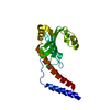
| ||||||||
|---|---|---|---|---|---|---|---|---|---|
| 1 | 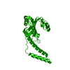
| ||||||||
| Unit cell |
| ||||||||
| Components on special symmetry positions |
|
-Components
| #1: Protein | Mass: 20548.670 Da / Num. of mol.: 1 / Fragment: UNP residues 46 to 227 Source method: isolated from a genetically manipulated source Source: (gene. exp.) Trypanosoma brucei (eukaryote) / Gene: Tb927.6.2420 / Plasmid: pET21a / Production host:  Escherichia coli (E. coli) / Strain (production host): Rosetta 2 / References: UniProt: Q584R4 Escherichia coli (E. coli) / Strain (production host): Rosetta 2 / References: UniProt: Q584R4 |
|---|---|
| #2: Water | ChemComp-HOH / Water Mass: 18.015 Da / Num. of mol.: 54 / Source method: isolated from a natural source / Formula: H2O Mass: 18.015 Da / Num. of mol.: 54 / Source method: isolated from a natural source / Formula: H2O |
-Experimental details
-Experiment
| Experiment | Method: X-RAY DIFFRACTION / Number of used crystals: 1 |
|---|
- Sample preparation
| Crystal | Density Matthews: 2.46 Å3/Da / Density % sol: 49.91 % |
|---|---|
| Crystal grow | Temperature: 298 K / Method: vapor diffusion, hanging drop / pH: 8 Details: 40% PEG400 0.1M Imidazole, pH 8, VAPOR DIFFUSION, HANGING DROP, temperature 298K |
-Data collection
| Diffraction | Mean temperature: 100 K |
|---|---|
| Diffraction source | Source: ROTATING ANODE / Type: RIGAKU RU300 / Wavelength: 1.5418 Å |
| Detector | Type: RIGAKU RAXIS IV / Detector: IMAGE PLATE / Date: 2009 / Details: mirrors |
| Radiation | Monochromator: mirrors / Protocol: SINGLE WAVELENGTH / Monochromatic (M) / Laue (L): M / Scattering type: x-ray |
| Radiation wavelength | Wavelength: 1.5418 Å / Relative weight: 1 |
| Reflection | Resolution: 2→29.32 Å / Num. all: 13481 / Num. obs: 13469 / % possible obs: 99.1 % / Observed criterion σ(F): 4.32 / Observed criterion σ(I): 5.48 / Biso Wilson estimate: 41.7 Å2 / Rmerge(I) obs: 0.083 |
| Reflection shell | Resolution: 2→2.11 Å / % possible all: 98.5 |
- Processing
| Software |
| |||||||||||||||||||||||||
|---|---|---|---|---|---|---|---|---|---|---|---|---|---|---|---|---|---|---|---|---|---|---|---|---|---|---|
| Refinement | Method to determine structure: MOLECULAR REPLACEMENT / Resolution: 2→29.3 Å / σ(F): 0 / Stereochemistry target values: CNS
| |||||||||||||||||||||||||
| Displacement parameters | Biso mean: 45.4776 Å2
| |||||||||||||||||||||||||
| Refinement step | Cycle: LAST / Resolution: 2→29.3 Å
| |||||||||||||||||||||||||
| Refine LS restraints |
|
