 Movie
Movie Controller

[English] 日本語
 Yorodumi
- PDB-3jsz: Legionella pneumophila glucosyltransferase Lgt1 N293A with UDP-Glc -
+ Open data

- Basic information
| Entry | Database: PDB / ID: 3jsz | ||||||
|---|---|---|---|---|---|---|---|
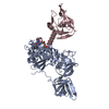
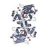
| Title | Legionella pneumophila glucosyltransferase Lgt1 N293A with UDP-Glc | ||||||
 Components Components | Putative uncharacterized protein | ||||||
 Keywords Keywords |  TRANSFERASE / glucosyltransferase / Legionnaire's disease / Legionella pneumophila TRANSFERASE / glucosyltransferase / Legionnaire's disease / Legionella pneumophila | ||||||
| Function / homology |  Function and homology information Function and homology information | ||||||
| Biological species |   Legionella pneumophila (bacteria) Legionella pneumophila (bacteria) | ||||||
| Method | X-RAY DIFFRACTION / SYNCHROTRON / MAD / Resolution: 1.7 Å | ||||||
 Authors Authors | Lu, W. / Du, J. / Belyi, Y. / Stahl, M. / Zivilikidis, T. / Gerhardt, S. / Aktories, K. / Einsle, O. | ||||||
 Citation Citation | Journal: J.Mol.Biol. / Year: 2009 Title: Structural Basis of the Action of Glucosyltransferase Lgt1 from Legionella pneumophila. Authors: Lu, W. / Du, J. / Stahl, M. / Tzivelekidis, T. / Belyi, Y. / Gerhardt, S. / Aktories, K. / Einsle, O. | ||||||
| History |
|
- Structure visualization
| Structure viewer | Molecule: MolmilJmol/JSmol |
|---|
- Downloads & links
-Download
| PDBx/mmCIF format | 3jsz.cif.gz | 133.2 KB | Display | PDBx/mmCIF format |
|---|---|---|---|---|
| PDB format | pdb3jsz.ent.gz | 107.5 KB | Display | PDB format |
| PDBx/mmJSON format | 3jsz.json.gz | Tree view | PDBx/mmJSON format | |
| Others |  Other downloads Other downloads |
-Validation report
| Arichive directory | https://data.pdbj.org/pub/pdb/validation_reports/js/3jszftp://data.pdbj.org/pub/pdb/validation_reports/js/3jsz | HTTPS FTP |
|---|
-Related structure data


-Links
 PDBj
PDBj
- Assembly
| Deposited unit | 
| ||||||||
|---|---|---|---|---|---|---|---|---|---|
| 1 |
| ||||||||
| Unit cell |
|
-Components
| #1: Protein | Mass: 60100.418 Da / Num. of mol.: 1 / Mutation: N293A / Source method: isolated from a natural source / Source: (natural) Legionella pneumophila (bacteria) / Strain: Lens / References: UniProt: Q5WWY0 |
|---|---|
| #2: Chemical | ChemComp-UPG / Uridine diphosphate glucose  Mass: 566.302 Da / Num. of mol.: 1 / Source method: obtained synthetically / Formula: C15H24N2O17P2 Mass: 566.302 Da / Num. of mol.: 1 / Source method: obtained synthetically / Formula: C15H24N2O17P2 |
| #3: Chemical | ChemComp-MG /   Mass: 24.305 Da / Num. of mol.: 1 / Source method: obtained synthetically / Formula: Mg Mass: 24.305 Da / Num. of mol.: 1 / Source method: obtained synthetically / Formula: Mg |
| #4: Water | ChemComp-HOH / Water Mass: 18.015 Da / Num. of mol.: 712 / Source method: isolated from a natural source / Formula: H2O Mass: 18.015 Da / Num. of mol.: 712 / Source method: isolated from a natural source / Formula: H2O |
-Experimental details
-Experiment
| Experiment | Method: X-RAY DIFFRACTION / Number of used crystals: 1 |
|---|
- Sample preparation
| Crystal | Density Matthews: 2.46 Å3/Da / Density % sol: 49.97 % |
|---|---|
| Crystal grow | Temperature: 293.15 K / pH: 5 Details: 23% PEG3350, 0.06M ammonium acetat, 0.1M sodium acetate buffer, pH5.0, temperature 293.15K |
-Data collection
| Diffraction source | Source: SYNCHROTRON / Site: SLS  / Beamline: X06SA / Wavelength: 1.00, 0.97973, 0.97957, 0.97205 / Beamline: X06SA / Wavelength: 1.00, 0.97973, 0.97957, 0.97205 | |||||||||||||||
|---|---|---|---|---|---|---|---|---|---|---|---|---|---|---|---|---|
| Detector | Type: PSI PILATUS 6M / Detector: PIXEL / Date: Jun 7, 2009 | |||||||||||||||
| Radiation | Protocol: MAD / Monochromatic (M) / Laue (L): M / Scattering type: x-ray | |||||||||||||||
| Radiation wavelength |
| |||||||||||||||
| Reflection | Resolution: 1.7→22 Å / Num. obs: 62915 / % possible obs: 99.9 % / Observed criterion σ(F): 2 / Observed criterion σ(I): 2 / Redundancy: 6.9 % / Rmerge(I) obs: 0.062 / Net I/σ(I): 7.3 | |||||||||||||||
| Reflection shell | Resolution: 1.7→1.8 Å / Redundancy: 6.6 % / Rmerge(I) obs: 0.391 / Mean I/σ(I) obs: 1.9 / Num. unique all: 60773 / % possible all: 100 |
- Processing
| Software | Name: REFMAC / Version: 5.5.0072 / Classification: refinement | ||||||||||||||||||||||||||||||||||||||||||||||||||||||||||||||||||||||||||||||||||||||||||||||||||||||||||||||||||||||||||||||||||||||||||||||||||||||||||||||||||||||||||
|---|---|---|---|---|---|---|---|---|---|---|---|---|---|---|---|---|---|---|---|---|---|---|---|---|---|---|---|---|---|---|---|---|---|---|---|---|---|---|---|---|---|---|---|---|---|---|---|---|---|---|---|---|---|---|---|---|---|---|---|---|---|---|---|---|---|---|---|---|---|---|---|---|---|---|---|---|---|---|---|---|---|---|---|---|---|---|---|---|---|---|---|---|---|---|---|---|---|---|---|---|---|---|---|---|---|---|---|---|---|---|---|---|---|---|---|---|---|---|---|---|---|---|---|---|---|---|---|---|---|---|---|---|---|---|---|---|---|---|---|---|---|---|---|---|---|---|---|---|---|---|---|---|---|---|---|---|---|---|---|---|---|---|---|---|---|---|---|---|---|---|---|
| Refinement | Method to determine structure: MAD / Resolution: 1.7→21.96 Å / Cor.coef. Fo:Fc: 0.965 / Cor.coef. Fo:Fc free: 0.955 / SU B: 3.903 / SU ML: 0.061 / TLS residual ADP flag: LIKELY RESIDUAL / Cross valid method: THROUGHOUT / ESU R: 0.101 / ESU R Free: 0.096 / Stereochemistry target values: MAXIMUM LIKELIHOOD / Details: HYDROGENS HAVE BEEN ADDED IN THE RIDING POSITIONS
| ||||||||||||||||||||||||||||||||||||||||||||||||||||||||||||||||||||||||||||||||||||||||||||||||||||||||||||||||||||||||||||||||||||||||||||||||||||||||||||||||||||||||||
| Solvent computation | Ion probe radii: 0.8 Å / Shrinkage radii: 0.8 Å / VDW probe radii: 1.4 Å / Solvent model: BABINET MODEL WITH MASK | ||||||||||||||||||||||||||||||||||||||||||||||||||||||||||||||||||||||||||||||||||||||||||||||||||||||||||||||||||||||||||||||||||||||||||||||||||||||||||||||||||||||||||
| Displacement parameters | Biso mean: 17.846 Å2
| ||||||||||||||||||||||||||||||||||||||||||||||||||||||||||||||||||||||||||||||||||||||||||||||||||||||||||||||||||||||||||||||||||||||||||||||||||||||||||||||||||||||||||
| Refinement step | Cycle: LAST / Resolution: 1.7→21.96 Å
| ||||||||||||||||||||||||||||||||||||||||||||||||||||||||||||||||||||||||||||||||||||||||||||||||||||||||||||||||||||||||||||||||||||||||||||||||||||||||||||||||||||||||||
| Refine LS restraints |
| ||||||||||||||||||||||||||||||||||||||||||||||||||||||||||||||||||||||||||||||||||||||||||||||||||||||||||||||||||||||||||||||||||||||||||||||||||||||||||||||||||||||||||
| LS refinement shell | Resolution: 1.7→1.744 Å / Total num. of bins used: 20
| ||||||||||||||||||||||||||||||||||||||||||||||||||||||||||||||||||||||||||||||||||||||||||||||||||||||||||||||||||||||||||||||||||||||||||||||||||||||||||||||||||||||||||
| Refinement TLS params. | Method: refined / Origin x: 32.6081 Å / Origin y: 4.4195 Å / Origin z: 1.2796 Å
|
