 Movie
Movie Controller

[English] 日本語
 Yorodumi
- PDB-3jsq: Crystal structure of adipocyte fatty acid binding protein non-cov... -
+ Open data

- Basic information
| Entry | Database: PDB / ID: 3jsq | ||||||
|---|---|---|---|---|---|---|---|
| Title | Crystal structure of adipocyte fatty acid binding protein non-covalently modified with 4-hydroxy-2-nonenal | ||||||
 Components Components | Adipocyte fatty acid-binding protein | ||||||
 Keywords Keywords | LIPID BINDING PROTEIN /  fatty acid binding protein fatty acid binding protein | ||||||
| Function / homology |  Function and homology information Function and homology informationTriglyceride catabolism / hormone receptor binding / long-chain fatty acid transmembrane transporter activity / cellular response to lithium ion / long-chain fatty acid binding / white fat cell differentiation / fatty acid transport / long-chain fatty acid transport / brown fat cell differentiation / fatty acid metabolic process ...Triglyceride catabolism / hormone receptor binding / long-chain fatty acid transmembrane transporter activity / cellular response to lithium ion / long-chain fatty acid binding / white fat cell differentiation / fatty acid transport / long-chain fatty acid transport / brown fat cell differentiation / fatty acid metabolic process / cholesterol homeostasis / fatty acid binding / response to bacterium / positive regulation of inflammatory response / cellular response to tumor necrosis factor / positive regulation of cold-induced thermogenesis / negative regulation of DNA-templated transcription / positive regulation of cell population proliferation / nucleoplasm / nucleus / cytosol / cytoplasmSimilarity search - Function | ||||||
| Biological species |  Mus musculus (house mouse) Mus musculus (house mouse) | ||||||
| Method | X-RAY DIFFRACTION / SYNCHROTRON / MOLECULAR REPLACEMENT / Resolution: 2.3 Å | ||||||
 Authors Authors | Hellberg, K. / Grimsrud, P.A. / Kruse, A.C. / Banaszak, L.J. / Ohlendorf, D.H. / Bernlohr, D.A. | ||||||
 Citation Citation | Journal: Protein Sci. / Year: 2010 Title: X-ray crystallographic analysis of adipocyte fatty acid binding protein (aP2) modified with 4-hydroxy-2-nonenal. Authors: Hellberg, K. / Grimsrud, P.A. / Kruse, A.C. / Banaszak, L.J. / Ohlendorf, D.H. / Bernlohr, D.A. | ||||||
| History |
|
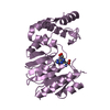
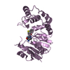
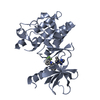
- Structure visualization
| Structure viewer | Molecule: MolmilJmol/JSmol |
|---|
- Downloads & links
-Download
| PDBx/mmCIF format | 3jsq.cif.gz | 41.9 KB | Display | PDBx/mmCIF format |
|---|---|---|---|---|
| PDB format | pdb3jsq.ent.gz | 28.6 KB | Display | PDB format |
| PDBx/mmJSON format | 3jsq.json.gz | Tree view | PDBx/mmJSON format | |
| Others |  Other downloads Other downloads |
-Validation report
| Arichive directory | https://data.pdbj.org/pub/pdb/validation_reports/js/3jsqftp://data.pdbj.org/pub/pdb/validation_reports/js/3jsq | HTTPS FTP |
|---|
-Related structure data
| Related structure data |  3js1C 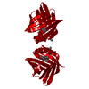 1lieS C: citing same article ( S: Starting model for refinement |
|---|---|
| Similar structure data |


-Links
 PDBj
PDBj

- Assembly
| Deposited unit | 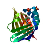
| ||||||||
|---|---|---|---|---|---|---|---|---|---|
| 1 | 
| ||||||||
| Unit cell |
| ||||||||
| Components on special symmetry positions |
| ||||||||
| Details | Authors state that the assembly is a dimer. The second part of the biological assembly is generated by the two fold axis: [-X,Y,1/2-Z] as shown in remark 350 |
-Components
| #1: Protein | Mass: 14539.688 Da / Num. of mol.: 1 Source method: isolated from a genetically manipulated source Source: (gene. exp.) Mus musculus (house mouse) / Gene: Fabp4, Ap2 / Plasmid: Modified pRSETb / Production host:  Escherichia coli (E. coli) / Strain (production host): BL21(DE3)pLysS / References: UniProt: P04117 Escherichia coli (E. coli) / Strain (production host): BL21(DE3)pLysS / References: UniProt: P04117 | ||
|---|---|---|---|
| #2: Chemical | ChemComp-CL / Chloride  Mass: 35.453 Da / Num. of mol.: 1 / Source method: obtained synthetically / Formula: Cl Mass: 35.453 Da / Num. of mol.: 1 / Source method: obtained synthetically / Formula: Cl | ||
| #3: Chemical | ChemComp-HNE / (  Mass: 156.222 Da / Num. of mol.: 1 / Source method: obtained synthetically / Formula: C9H16O2 Mass: 156.222 Da / Num. of mol.: 1 / Source method: obtained synthetically / Formula: C9H16O2 | ||
| #4: Chemical | ChemComp-PO4 / Phosphate  Mass: 94.971 Da / Num. of mol.: 4 / Source method: obtained synthetically / Formula: PO4 Mass: 94.971 Da / Num. of mol.: 4 / Source method: obtained synthetically / Formula: PO4#5: Water | ChemComp-HOH / | Water Mass: 18.015 Da / Num. of mol.: 91 / Source method: isolated from a natural source / Formula: H2O Mass: 18.015 Da / Num. of mol.: 91 / Source method: isolated from a natural source / Formula: H2O |
-Experimental details
-Experiment
| Experiment | Method: X-RAY DIFFRACTION / Number of used crystals: 1 |
|---|
- Sample preparation
| Crystal | Density Matthews: 3.05 Å3/Da / Density % sol: 59.72 % |
|---|---|
| Crystal grow | Temperature: 291 K / Method: vapor diffusion, sitting drop / pH: 7.5 Details: 0.1 M HEPES, 1.6 M Sodium/potassium phosphate, pH 7.5, VAPOR DIFFUSION, SITTING DROP, temperature 291K |
-Data collection
| Diffraction | Mean temperature: 100 K |
|---|---|
| Diffraction source | Source: SYNCHROTRON / Site: APS  / Beamline: 23-ID-B / Wavelength: 0.97934 Å / Beamline: 23-ID-B / Wavelength: 0.97934 Å |
| Detector | Type: MARMOSAIC 300 mm CCD / Detector: CCD / Date: Mar 29, 2008 / Details: Mirrors |
| Radiation | Monochromator: Si(111) / Protocol: SINGLE WAVELENGTH / Monochromatic (M) / Laue (L): M / Scattering type: x-ray |
| Radiation wavelength | Wavelength: 0.97934 Å / Relative weight: 1 |
| Reflection | Resolution: 2.3→50 Å / Num. all: 7758 / Num. obs: 7758 / % possible obs: 93.7 % / Redundancy: 3.6 % / Rsym value: 0.065 / Net I/σ(I): 16.63 |
| Reflection shell | Resolution: 2.3→2.34 Å / Redundancy: 3.2 % / Mean I/σ(I) obs: 2.85 / Num. unique all: 297 / Rsym value: 0.332 / % possible all: 73.5 |
- Processing
| Software |
| ||||||||||||||||||||||||||||||||||||||||||||||||||||||||||||||||||||||||||||||||||||||||||
|---|---|---|---|---|---|---|---|---|---|---|---|---|---|---|---|---|---|---|---|---|---|---|---|---|---|---|---|---|---|---|---|---|---|---|---|---|---|---|---|---|---|---|---|---|---|---|---|---|---|---|---|---|---|---|---|---|---|---|---|---|---|---|---|---|---|---|---|---|---|---|---|---|---|---|---|---|---|---|---|---|---|---|---|---|---|---|---|---|---|---|---|
| Refinement | Method to determine structure: MOLECULAR REPLACEMENT Starting model: PDB entry 1LIE Resolution: 2.3→19.97 Å / Cor.coef. Fo:Fc: 0.932 / Cor.coef. Fo:Fc free: 0.904 / Occupancy max: 1 / Occupancy min: 0.5 / Isotropic thermal model: Isotropic / Cross valid method: THROUGHOUT / σ(F): 0 / ESU R: 0.325 / ESU R Free: 0.247 / Stereochemistry target values: MAXIMUM LIKELIHOOD
| ||||||||||||||||||||||||||||||||||||||||||||||||||||||||||||||||||||||||||||||||||||||||||
| Solvent computation | Ion probe radii: 0.8 Å / Shrinkage radii: 0.8 Å / VDW probe radii: 1.2 Å / Solvent model: MASK | ||||||||||||||||||||||||||||||||||||||||||||||||||||||||||||||||||||||||||||||||||||||||||
| Displacement parameters | Biso max: 96.42 Å2 / Biso mean: 34.676 Å2 / Biso min: 16.97 Å2
| ||||||||||||||||||||||||||||||||||||||||||||||||||||||||||||||||||||||||||||||||||||||||||
| Refinement step | Cycle: LAST / Resolution: 2.3→19.97 Å
| ||||||||||||||||||||||||||||||||||||||||||||||||||||||||||||||||||||||||||||||||||||||||||
| Refine LS restraints |
| ||||||||||||||||||||||||||||||||||||||||||||||||||||||||||||||||||||||||||||||||||||||||||
| LS refinement shell | Resolution: 2.3→2.36 Å / Total num. of bins used: 20
|
