 Movie
Movie Controller


+ Open data

- Basic information
| Entry | Database: PDB / ID: 3jrz | ||||||
|---|---|---|---|---|---|---|---|
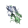
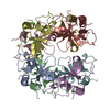
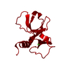
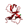
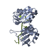
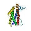
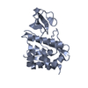
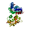
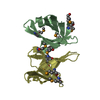
| Title | CcdBVfi-FormII-pH5.6 | ||||||
 Components Components | CcdB Christian Commission for Development in Bangladesh Christian Commission for Development in Bangladesh | ||||||
 Keywords Keywords | TOXIN / ALPHA+BETA / SH3 domain | ||||||
| Function / homology |  Function and homology information Function and homology informationDNA topoisomerase type II (double strand cut, ATP-hydrolyzing) inhibitor activity / plasmid maintenanceSimilarity search - Function | ||||||
| Biological species |  Vibrio fischeri (bacteria) Vibrio fischeri (bacteria) | ||||||
| Method | X-RAY DIFFRACTION / SYNCHROTRON / MOLECULAR REPLACEMENT / Resolution: 1.7 Å | ||||||
 Authors Authors | De Jonge, N. / Buts, L. / Loris, R. | ||||||
 Citation Citation | Journal: J.Biol.Chem. / Year: 2010 Title: Structural and thermodynamic characterization of vibrio fischeri CCDB Authors: De Jonge, N. / Hohlweg, W. / Garcia-Pino, A. / Respondek, M. / Buts, L. / Haesaerts, S. / Lah, J. / Zangger, K. / Loris, R. #1: Journal: Acta Crystallogr.,Sect.F / Year: 2007 Title: Purification and crystallization of Vibrio fischeri CcdB and its complexes with fragments of gyrase and CcdA Authors: De Jonge, N. / Buts, L. / Vangelooven, J. / Mine, N. / Van Melderen, L. / Wyns, L. / Loris, R. | ||||||
| History |
|
- Structure visualization
| Structure viewer | Molecule: MolmilJmol/JSmol |
|---|
- Downloads & links
-Download
| PDBx/mmCIF format | 3jrz.cif.gz | 33.2 KB | Display | PDBx/mmCIF format |
|---|---|---|---|---|
| PDB format | pdb3jrz.ent.gz | 21.8 KB | Display | PDB format |
| PDBx/mmJSON format | 3jrz.json.gz | Tree view | PDBx/mmJSON format | |
| Others |  Other downloads Other downloads |
-Validation report
| Arichive directory | https://data.pdbj.org/pub/pdb/validation_reports/jr/3jrzftp://data.pdbj.org/pub/pdb/validation_reports/jr/3jrz | HTTPS FTP |
|---|
-Related structure data
| Related structure data | 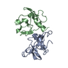 2kmtC 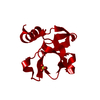 3jscSC S: Starting model for refinement C: citing same article ( |
|---|---|
| Similar structure data |


-Links
 PDBj
PDBj- Assembly
| Deposited unit | 
| ||||||||
|---|---|---|---|---|---|---|---|---|---|
| 1 | 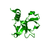
| ||||||||
| Unit cell |
|
-Components
| #1: Protein | Christian Commission for Development in Bangladesh Mass: 11879.721 Da / Num. of mol.: 1 Source method: isolated from a genetically manipulated source Details: tac promotor / Source: (gene. exp.) Vibrio fischeri (bacteria) / Gene: ccdB / Plasmid: pKK223-3 / Production host: Escherichia coli (E. coli) / Strain (production host): B462 / References: UniProt: Q84B82 |
|---|---|
| #2: Water | ChemComp-HOH / Water Mass: 18.015 Da / Num. of mol.: 73 / Source method: isolated from a natural source / Formula: H2O Mass: 18.015 Da / Num. of mol.: 73 / Source method: isolated from a natural source / Formula: H2O |
-Experimental details
-Experiment
| Experiment | Method: X-RAY DIFFRACTION / Number of used crystals: 1 |
|---|
- Sample preparation
| Crystal | Density Matthews: 1.89 Å3/Da / Density % sol: 34.99 % |
|---|---|
| Crystal grow | Temperature: 293 K / Method: vapor diffusion, hanging drop / pH: 5.6 Details: 100mM sodium citrate pH5.6, 2% polyethyleneimine, 500mM NaCl, VAPOR DIFFUSION, HANGING DROP, temperature 293K |
-Data collection
| Diffraction | Mean temperature: 100 K |
|---|---|
| Diffraction source | Source: SYNCHROTRON / Site: EMBL/DESY, HAMBURG  / Beamline: X11 / Wavelength: 0.8162 Å / Beamline: X11 / Wavelength: 0.8162 Å |
| Detector | Type: MAR CCD 165 mm / Detector: CCD / Date: Oct 12, 2006 |
| Radiation | Protocol: SINGLE WAVELENGTH / Monochromatic (M) / Laue (L): M / Scattering type: x-ray |
| Radiation wavelength | Wavelength: 0.8162 Å / Relative weight: 1 |
| Reflection | Resolution: 1.7→19.9 Å / Num. all: 9956 / Num. obs: 9956 / % possible obs: 99.9 % / Observed criterion σ(F): 0 / Observed criterion σ(I): -3 / Redundancy: 6.31 % / Biso Wilson estimate: 17.46 Å2 / Rmerge(I) obs: 0.04 / Rsym value: 0.04 / Net I/σ(I): 22.98 |
| Reflection shell | Resolution: 1.7→1.76 Å / Redundancy: 6.3 % / Rmerge(I) obs: 0.237 / Mean I/σ(I) obs: 8 / Num. unique all: 986 / Rsym value: 0.237 / % possible all: 100 |
- Processing
| Software |
| ||||||||||||||||||||||||||||
|---|---|---|---|---|---|---|---|---|---|---|---|---|---|---|---|---|---|---|---|---|---|---|---|---|---|---|---|---|---|
| Refinement | Method to determine structure: MOLECULAR REPLACEMENT Starting model: PDB ENTRY 3JSC Resolution: 1.7→18.776 Å / Occupancy max: 1 / Occupancy min: 0.49 / FOM work R set: 0.879 / SU ML: 0.22 / Isotropic thermal model: individual atomic B factors / Cross valid method: THROUGHOUT / σ(F): 1.36 / σ(I): 0 / Phase error: 18.79 / Stereochemistry target values: ML
| ||||||||||||||||||||||||||||
| Solvent computation | Shrinkage radii: 0.9 Å / VDW probe radii: 1.11 Å / Solvent model: FLAT BULK SOLVENT MODEL / Bsol: 72.977 Å2 / ksol: 0.414 e/Å3 | ||||||||||||||||||||||||||||
| Displacement parameters | Biso max: 45.63 Å2 / Biso mean: 18.774 Å2 / Biso min: 9.33 Å2
| ||||||||||||||||||||||||||||
| Refinement step | Cycle: LAST / Resolution: 1.7→18.776 Å
| ||||||||||||||||||||||||||||
| Refine LS restraints |
| ||||||||||||||||||||||||||||
| LS refinement shell |
|
