Movie
Movie Controller

[English] 日本語
 Yorodumi
- PDB-3jbq: Domain Organization and Conformational Plasticity of the G Protei... -
+ Open data

- Basic information
| Entry | Database: PDB / ID: 3jbq | ||||||
|---|---|---|---|---|---|---|---|
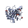
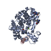
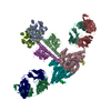
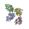
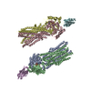
| Title | Domain Organization and Conformational Plasticity of the G Protein Effector, PDE6 | ||||||
 Components Components |
| ||||||
 Keywords Keywords | HYDROLASE/IMMUNE SYSTEM / Phosphodiesterase / photoreceptor / PDE6 / HYDROLASE-IMMUNE SYSTEM complex | ||||||
| Function / homology |  Function and homology information Function and homology informationcGMP effects / Smooth Muscle Contraction / RHOBTB1 GTPase cycle / cyclic-nucleotide phosphodiesterase activity / GMP catabolic process / cellular response to macrophage colony-stimulating factor stimulus / 3',5'-cyclic-GMP phosphodiesterase / cellular response to cGMP / Inactivation, recovery and regulation of the phototransduction cascade / negative regulation of cAMP-mediated signaling ...cGMP effects / Smooth Muscle Contraction / RHOBTB1 GTPase cycle / cyclic-nucleotide phosphodiesterase activity / GMP catabolic process / cellular response to macrophage colony-stimulating factor stimulus / 3',5'-cyclic-GMP phosphodiesterase / cellular response to cGMP / Inactivation, recovery and regulation of the phototransduction cascade / negative regulation of cAMP-mediated signaling / positive regulation of G protein-coupled receptor signaling pathway / Activation of the phototransduction cascade / positive regulation of vascular permeability / cellular response to granulocyte macrophage colony-stimulating factor stimulus / negative regulation of vascular permeability / establishment of endothelial barrier / regulation of mitochondrion organization / Ca2+ pathway / positive regulation of epidermal growth factor receptor signaling pathway / photoreceptor outer segment membrane / 3',5'-cGMP-stimulated cyclic-nucleotide phosphodiesterase activity / 3',5'-cyclic-nucleotide phosphodiesterase / negative regulation of cGMP-mediated signaling / cGMP-mediated signaling / cGMP catabolic process / cGMP binding / 3',5'-cyclic-GMP phosphodiesterase activity / 3',5'-cyclic-AMP phosphodiesterase activity / regulation of cAMP-mediated signaling / visual perception / cAMP-mediated signaling / synaptic membrane / cellular response to mechanical stimulus / photoreceptor disc membrane / positive regulation of inflammatory response / presynaptic membrane / mitochondrial outer membrane / molecular adaptor activity / mitochondrial inner membrane / mitochondrial matrix / positive regulation of gene expression / perinuclear region of cytoplasm / negative regulation of transcription by RNA polymerase II / Golgi apparatus / signal transduction / endoplasmic reticulum / protein homodimerization activity / zinc ion binding / nucleus / metal ion binding / plasma membrane / cytoplasm / cytosol Similarity search - Function | ||||||
| Biological species |  | ||||||
| Method | ELECTRON MICROSCOPY / single particle reconstruction / cryo EM / Resolution: 11 Å | ||||||
 Authors Authors | Zhang, Z. / He, F. / Constantine, R. / Baker, M.L. / Baehr, W. / Schmid, M.F. / Wensel, T.G. / Agosto, M.A. | ||||||
 Citation Citation | Journal: J Biol Chem / Year: 2015 Title: Domain organization and conformational plasticity of the G protein effector, PDE6. Authors: Zhixian Zhang / Feng He / Ryan Constantine / Matthew L Baker / Wolfgang Baehr / Michael F Schmid / Theodore G Wensel / Melina A Agosto /  Abstract: The cGMP phosphodiesterase of rod photoreceptor cells, PDE6, is the key effector enzyme in phototransduction. Two large catalytic subunits, PDE6α and -β, each contain one catalytic domain and two ...The cGMP phosphodiesterase of rod photoreceptor cells, PDE6, is the key effector enzyme in phototransduction. Two large catalytic subunits, PDE6α and -β, each contain one catalytic domain and two non-catalytic GAF domains, whereas two small inhibitory PDE6γ subunits allow tight regulation by the G protein transducin. The structure of holo-PDE6 in complex with the ROS-1 antibody Fab fragment was determined by cryo-electron microscopy. The ∼11 Å map revealed previously unseen features of PDE6, and each domain was readily fit with high resolution structures. A structure of PDE6 in complex with prenyl-binding protein (PrBP/δ) indicated the location of the PDE6 C-terminal prenylations. Reconstructions of complexes with Fab fragments bound to N or C termini of PDE6γ revealed that PDE6γ stretches from the catalytic domain at one end of the holoenzyme to the GAF-A domain at the other. Removal of PDE6γ caused dramatic structural rearrangements, which were reversed upon its restoration. | ||||||
| History |
|
- Structure visualization
| Movie |
Movie viewer |
|---|---|
| Structure viewer | Molecule: MolmilJmol/JSmol |
- Downloads & links
-Download
| PDBx/mmCIF format | 3jbq.cif.gz | 428.3 KB | Display | PDBx/mmCIF format |
|---|---|---|---|---|
| PDB format | pdb3jbq.ent.gz | 357.2 KB | Display | PDB format |
| PDBx/mmJSON format | 3jbq.json.gz | Tree view | PDBx/mmJSON format | |
| Others |  Other downloads Other downloads |
-Validation report
| Summary document | 3jbq_validation.pdf.gz | 666.4 KB | Display | wwPDB validaton report |
|---|---|---|---|---|
| Full document | 3jbq_full_validation.pdf.gz | 671.4 KB | Display | |
| Data in XML | 3jbq_validation.xml.gz | 54.2 KB | Display | |
| Data in CIF | 3jbq_validation.cif.gz | 85.5 KB | Display | |
| Arichive directory | https://data.pdbj.org/pub/pdb/validation_reports/jb/3jbqftp://data.pdbj.org/pub/pdb/validation_reports/jb/3jbq | HTTPS FTP |
-Related structure data
| Related structure data |  6258MC  3jabC M: map data used to model this data C: citing same article ( |
|---|---|
| Similar structure data |


-Links
 PDBj
PDBj


- Assembly
| Deposited unit | 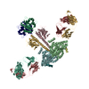
|
|---|---|
| 1 |
|
-Components
-Phosphodiesterase ... , 2 types, 4 molecules BFDX
| #3: Protein | Mass: 38282.086 Da / Num. of mol.: 2 / Fragment: SEE REMARK 999 / Source method: isolated from a natural source / Source: (natural) #4: Protein/peptide | Mass: 2185.436 Da / Num. of mol.: 2 / Fragment: UNP residues 70-87 / Source method: isolated from a natural source / Source: (natural) |
|---|
-Protein , 2 types, 4 molecules CG12
| #5: Protein | Mass: 20671.434 Da / Num. of mol.: 2 / Fragment: SEE REMARK 999 / Source method: isolated from a natural source / Source: (natural) #6: Protein | Mass: 21554.822 Da / Num. of mol.: 2 / Fragment: SEE REMARK 999 / Source method: isolated from a natural source / Source: (natural) |
|---|
-Antibody , 2 types, 4 molecules LlHh
| #1: Antibody | Mass: 23540.990 Da / Num. of mol.: 2 / Fragment: Fab / Source method: isolated from a natural source / Source: (natural) #2: Antibody | Mass: 23781.471 Da / Num. of mol.: 2 / Fragment: Fab / Source method: isolated from a natural source / Source: (natural) |
|---|
-Details
| Sequence details | THE IMAGED PDE6 WAS DERIVED FROM BOS TAURUS, BUT SOME MODELED SEQUENCES ARE FROM HOMO SAPIENS ...THE IMAGED PDE6 WAS DERIVED FROM BOS TAURUS, BUT SOME MODELED SEQUENCES ARE FROM HOMO SAPIENS (CHAINS B, C, F, G) OR GALLUS GALLUS (CHAINS 1, 2). THE PHOSPHODIE |
|---|
-Experimental details
-Experiment
| Experiment | Method: ELECTRON MICROSCOPY |
|---|---|
| EM experiment | Aggregation state: PARTICLE / 3D reconstruction method: single particle reconstruction |
- Sample preparation
| Component |
| ||||||||||||||||||||
|---|---|---|---|---|---|---|---|---|---|---|---|---|---|---|---|---|---|---|---|---|---|
| Molecular weight | Value: 0.32 MDa / Experimental value: NO | ||||||||||||||||||||
| Buffer solution | Name: 20 mM sodium phosphate, 150 mM sodium chloride / pH: 7.5 / Details: 20 mM sodium phosphate, 150 mM sodium chloride | ||||||||||||||||||||
| Specimen | Conc.: 0.5 mg/ml / Embedding applied: NO / Shadowing applied: NO / Staining applied: NO / Vitrification applied: YES | ||||||||||||||||||||
| Specimen support | Details: 400 mesh glow-discharged Quantifoil grids with 2.0 Angstrom holes | ||||||||||||||||||||
| Vitrification | Instrument: FEI VITROBOT MARK III / Cryogen name: ETHANE / Temp: 93 K / Humidity: 95 % Details: Applied 3 uL of sample per grid and blotted for 1 second before plunging into liquid ethane (FEI VITROBOT MARK III). Method: Applied 3 ul of sample per grid, blotted for 1 second before plunging |
- Electron microscopy imaging
| Microscopy | Model: JEOL 2010F / Date: Aug 8, 2011 / Details: Parallel beam illumination |
|---|---|
| Electron gun | Electron source:  FIELD EMISSION GUN / Accelerating voltage: 200 kV / Illumination mode: FLOOD BEAM FIELD EMISSION GUN / Accelerating voltage: 200 kV / Illumination mode: FLOOD BEAM |
| Electron lens | Mode: BRIGHT FIELD / Nominal magnification: 60000 X / Cs: 2 mm Astigmatism: Objective lens astigmatism was corrected at 100,000 times magnification. Camera length: 0 mm |
| Specimen holder | Specimen holder model: GATAN LIQUID NITROGEN / Temperature (max): 94 K |
| Image recording | Electron dose: 15 e/Å2 / Film or detector model: GATAN ULTRASCAN 4000 (4k x 4k) |
| EM imaging optics | Energyfilter name: FEI |
| Radiation | Protocol: SINGLE WAVELENGTH / Monochromatic (M) / Laue (L): M / Scattering type: x-ray |
| Radiation wavelength | Relative weight: 1 |
- Processing
| EM software |
| ||||||||||||||||||||||||||||
|---|---|---|---|---|---|---|---|---|---|---|---|---|---|---|---|---|---|---|---|---|---|---|---|---|---|---|---|---|---|
| CTF correction | Details: by particle using CtFit | ||||||||||||||||||||||||||||
| Symmetry | Point symmetry: C2 (2 fold cyclic) | ||||||||||||||||||||||||||||
| 3D reconstruction | Method: projection matching / Resolution: 11 Å / Resolution method: FSC 0.143 CUT-OFF / Num. of particles: 12373 / Nominal pixel size: 1.81 Å / Actual pixel size: 1.81 Å Details: A total of 21100 particles were picked from ice images and CTF-corrected using Ctfit. Initial three-dimensional models were generated from either reference-free class averages or from a ...Details: A total of 21100 particles were picked from ice images and CTF-corrected using Ctfit. Initial three-dimensional models were generated from either reference-free class averages or from a cylinder. The two models were essentially similar. Three noise-seeded models were generated and used as initial models in Multirefine. A model with two Ros-1 Fabs bound emerged from a population of 15000 particles and was subjected to further refinement using standard iterative projection matching, class averaging, and Fourier reconstruction. The final three-dimensional map with C2 symmetry was generated from 12373 particles. Num. of class averages: 20 / Symmetry type: POINT | ||||||||||||||||||||||||||||
| Atomic model building | Protocol: RIGID BODY FIT / Space: REAL Details: REFINEMENT PROTOCOL--rigid body refinement DETAILS--The domains were fit using Chimera. The model was refined using Phenix real-space refinement. | ||||||||||||||||||||||||||||
| Atomic model building |
| ||||||||||||||||||||||||||||
| Refinement step | Cycle: LAST
|