 Movie
Movie Controller

[English] 日本語
 Yorodumi
- PDB-3jb8: Insight into Three-dimensional structure of Maize Chlorotic Mottl... -
+ Open data

- Basic information
| Entry | Database: PDB / ID: 3jb8 | ||||||
|---|---|---|---|---|---|---|---|
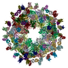
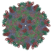
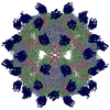
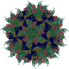
| Title | Insight into Three-dimensional structure of Maize Chlorotic Mottle Virus Revealed by Single Particle Analysis | ||||||
 Components Components | Coat protein | ||||||
 Keywords Keywords |  VIRUS / MCMV / Single Particle Analysis / Three-dimensional Structure / icosahedral virus / Transmission VIRUS / MCMV / Single Particle Analysis / Three-dimensional Structure / icosahedral virus / Transmission | ||||||
| Function / homology |  Function and homology information Function and homology information | ||||||
| Biological species |  Maize chlorotic mottle virus Maize chlorotic mottle virus | ||||||
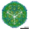
| Method | ELECTRON MICROSCOPY / single particle reconstruction / cryo EM / Resolution: 3.6 Å | ||||||
 Authors Authors | Wang, C.Y. / Zhang, Q.F. / Gao, Y.Z. / Zhou, X.P. / Ji, G. / Huang, X.J. / Hong, J. / Zhang, C.X. | ||||||
 Citation Citation | Journal: Virology / Year: 2015 Title: Insight into the three-dimensional structure of maize chlorotic mottle virus revealed by Cryo-EM single particle analysis. Authors: Chun-Yan Wang / Qin-Fen Zhang / Yuan-Zhu Gao / Xue-Ping Zhou / Gang Ji / Xiao-Jun Huang / Jian Hong / Chuan-Xi Zhang /  Abstract: Maize chlorotic mottle virus (MCMV) is the only member of the Machlomovirus genus in the family Tombusviridae. Here, we obtained the Cryo-EM structure of MCMV by single particle analysis with most ...Maize chlorotic mottle virus (MCMV) is the only member of the Machlomovirus genus in the family Tombusviridae. Here, we obtained the Cryo-EM structure of MCMV by single particle analysis with most local resolution at approximately 4 Å. The Cα backbone was built based on residues with bulky side chains. The resolved C-terminus of the capsid protein subunit and obvious openings at the 2-fold axis demonstrated the compactness of the asymmetric unit, which indicates an important role in the stability of MCMV. The Asp116 residue from each subunit around the 5-fold and 3-fold axes contributed to the negative charges in the centers of the pentamers and hexamers, which might serve as a solid barrier against the leakage of genomic RNA. Finally, the loops most exposed on the surface were analyzed and are proposed to be potential functional sites related to MCMV transmission. | ||||||
| History |
|
- Structure visualization
| Movie |
Movie viewer |
|---|---|
| Structure viewer | Molecule: MolmilJmol/JSmol |
- Downloads & links
-Download
| PDBx/mmCIF format | 3jb8.cif.gz | 102.9 KB | Display | PDBx/mmCIF format |
|---|---|---|---|---|
| PDB format | pdb3jb8.ent.gz | 81.5 KB | Display | PDB format |
| PDBx/mmJSON format | 3jb8.json.gz | Tree view | PDBx/mmJSON format | |
| Others |  Other downloads Other downloads |
-Validation report
| Arichive directory | https://data.pdbj.org/pub/pdb/validation_reports/jb/3jb8ftp://data.pdbj.org/pub/pdb/validation_reports/jb/3jb8 | HTTPS FTP |
|---|
-Related structure data
| Related structure data | 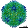 6382MC M: map data used to model this data C: citing same article ( |
|---|---|
| Similar structure data |
-Links
 PDBj
PDBj

- Assembly
| Deposited unit | 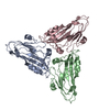
|
|---|---|
| 1 | x 60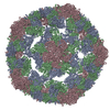
|
| 2 |
|
| 3 | x 5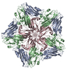
|
| 4 | x 6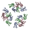
|
| 5 | 
|
| Symmetry | Point symmetry: (Schoenflies symbol: I (icosahedral)) |
-Components
| #1: Protein | Mass: 19654.631 Da / Num. of mol.: 3 / Fragment: UNP RESIDUES 45-229 / Source method: isolated from a natural source / Source: (natural) Maize chlorotic mottle virus / References: UniProt: I6QQC4 |
|---|
-Experimental details
-Experiment
| Experiment | Method: ELECTRON MICROSCOPY |
|---|---|
| EM experiment | Aggregation state: PARTICLE / 3D reconstruction method: single particle reconstruction |
- Sample preparation
| Component | Name: Maize chloroltic mottle virus / Type: VIRUS |
|---|---|
| Details of virus | Host category: PLANT / Type: VIRION |
| Buffer solution | Name: PBS / pH: 6 / Details: PBS |
| Specimen | Embedding applied: NO / Shadowing applied: NO / Staining applied: NO / Vitrification applied: YES |
| Specimen support | Details: This grid plus sample was kept at liquid nitrogen. |
| Vitrification | Instrument: FEI VITROBOT MARK IV / Cryogen name: NITROGEN / Humidity: 100 % / Method: blot for 2 seconds before plunging |
- Electron microscopy imaging
| Experimental equipment |  Model: Titan Krios / Image courtesy: FEI Company |
|---|---|
| Microscopy | Model: FEI TITAN KRIOS / Date: Oct 20, 2012 |
| Electron gun | Electron source: FIELD EMISSION GUN / Accelerating voltage: 300 kV / Illumination mode: FLOOD BEAM |
| Electron lens | Mode: BRIGHT FIELDBright-field microscopy / Nominal defocus max: 3000 nm / Nominal defocus min: 1000 nm / Cs: 2.7 mm / Camera length: 0 mm |
| Specimen holder | Specimen holder model: FEI TITAN KRIOS AUTOGRID HOLDER / Tilt angle max: 0 ° / Tilt angle min: 0 ° |
| Image recording | Film or detector model: GATAN ULTRASCAN 4000 (4k x 4k) |
- Processing
| EM software |
| ||||||||||||
|---|---|---|---|---|---|---|---|---|---|---|---|---|---|
| Symmetry | Point symmetry: I (icosahedral) | ||||||||||||
| 3D reconstruction | Resolution: 3.6 Å / Resolution method: FSC 0.143 CUT-OFF / Num. of particles: 53600 / Symmetry type: POINT | ||||||||||||
| Atomic model building | B value: 150 / Protocol: RIGID BODY FIT / Space: REAL / Target criteria: Cross-correlation coefficient / Details: REFINEMENT PROTOCOL--rigid body | ||||||||||||
| Refinement step | Cycle: LAST
|
