- PDB-3j6r: Electron cryo-microscopy of Human Papillomavirus Type 16 capsid -
+
Open data
ID or keywords:
Loading...
-
Basic information
Entry
Database: PDB / ID: 3j6r
Title
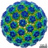
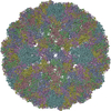
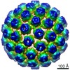
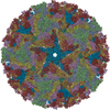
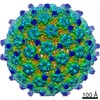
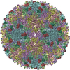
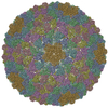
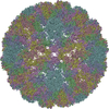
Electron cryo-microscopy of Human Papillomavirus Type 16 capsid
Components
Major capsid protein L1
Keywords
VIRUS / capsid protein
Function / homology
Function and homology information
T=7 icosahedral viral capsid / endocytosis involved in viral entry into host cell / host cell nucleus / structural molecule activity / virion attachment to host cell Similarity search - Function
Major capsid L1 (late) protein, Papillomavirus / Major capsid L1 (late) superfamily, Papillomavirus / L1 (late) protein / Double-stranded DNA virus, group I, capsid Similarity search - Domain/homology
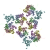
Journal: mBio / Year: 2014 Title: Maturation of the human papillomavirus 16 capsid. Authors: Giovanni Cardone / Adam L Moyer / Naiqian Cheng / Cynthia D Thompson / Israel Dvoretzky / Douglas R Lowy / John T Schiller / Alasdair C Steven / Christopher B Buck / Benes L Trus / Abstract: Papillomaviruses are a family of nonenveloped DNA viruses that infect the skin or mucosa of their vertebrate hosts. The viral life cycle is closely tied to the differentiation of infected ...Papillomaviruses are a family of nonenveloped DNA viruses that infect the skin or mucosa of their vertebrate hosts. The viral life cycle is closely tied to the differentiation of infected keratinocytes. Papillomavirus virions are released into the environment through a process known as desquamation, in which keratinocytes lose structural integrity prior to being shed from the surface of the skin. During this process, virions are exposed to an increasingly oxidative environment, leading to their stabilization through the formation of disulfide cross-links between neighboring molecules of the major capsid protein, L1. We used time-lapse cryo-electron microscopy and image analysis to study the maturation of HPV16 capsids assembled in mammalian cells and exposed to an oxidizing environment after cell lysis. Initially, the virion is a loosely connected procapsid that, under in vitro conditions, condenses over several hours into the more familiar 60-nm-diameter papillomavirus capsid. In this process, the procapsid shrinks by ~5% in diameter, its pentameric capsomers change in structure (most markedly in the axial region), and the interaction surfaces between adjacent capsomers are consolidated. A C175S mutant that cannot achieve normal inter-L1 disulfide cross-links shows maturation-related shrinkage but does not achieve the fully condensed 60-nm form. Pseudoatomic modeling based on a 9-Å resolution reconstruction of fully mature capsids revealed C-terminal disulfide-stabilized "suspended bridges" that form intercapsomeric cross-links. The data suggest a model in which procapsids exist in a range of dynamic intermediates that can be locked into increasingly mature configurations by disulfide cross-linking, possibly through a Brownian ratchet mechanism. Importance: Human papillomaviruses (HPVs) cause nearly all cases of cervical cancer, a major fraction of cancers of the penis, vagina/vulva, anus, and tonsils, and genital and nongenital warts. HPV types associated with a high risk of cancer, such as HPV16, are generally transmitted via sexual contact. The nonenveloped virion of HPVs shows a high degree of stability, allowing the virus to persist in an infectious form in environmental fomites. In this study, we used cryo-electron microscopy to elucidate the structure of the HPV16 capsid at different stages of maturation. The fully mature capsid adopts a rigid, highly regular structure stabilized by intermolecular disulfide bonds. The availability of a pseudoatomic model of the fully mature HPV16 virion should help guide understanding of antibody responses elicited by HPV capsid-based vaccines.
#200 - Aug 2016 Quasisymmetry in Icosahedral Viruses similarity (2)
-
Assembly
Deposited unit
B: Major capsid protein L1 A: Major capsid protein L1 E: Major capsid protein L1 D: Major capsid protein L1 C: Major capsid protein L1 F: Major capsid protein L1
B: Major capsid protein L1 A: Major capsid protein L1 E: Major capsid protein L1 D: Major capsid protein L1 C: Major capsid protein L1 F: Major capsid protein L1
B: Major capsid protein L1 A: Major capsid protein L1 E: Major capsid protein L1 D: Major capsid protein L1 C: Major capsid protein L1 F: Major capsid protein L1
x 5
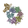
icosahedral pentamer
1.6 MDa, 30 polymers
Theoretical mass
Number of molelcules
Total (without water)
1,603,369
30
Polymers
1,603,369
30
Non-polymers
0
0
Water
0
Type
Name
Symmetry operation
Number
identity operation
1_555
x,y,z
1
point symmetry operation
4
4
B: Major capsid protein L1 A: Major capsid protein L1 E: Major capsid protein L1 D: Major capsid protein L1 C: Major capsid protein L1 F: Major capsid protein L1
x 6
icosahedral 23 hexamer
1.92 MDa, 36 polymers
Theoretical mass
Number of molelcules
Total (without water)
1,924,042
36
Polymers
1,924,042
36
Non-polymers
0
0
Water
0
Type
Name
Symmetry operation
Number
identity operation
1_555
x,y,z
1
point symmetry operation
5
5
Idetical with deposited unit in distinct coordinate
icosahedral asymmetric unit, std point frame
Type
Name
Symmetry operation
Number
transform to point frame
1
Symmetry
Point symmetry: (Schoenflies symbol: I (icosahedral))
-
Components
#1: Protein
MajorcapsidproteinL1 / L1 protein
Mass: 53445.617 Da / Num. of mol.: 6 / Fragment: UNP residues 35-512 / Source method: isolated from a natural source / Source: (natural) Human papillomavirus type 16 / References: UniProt: Q4VRM0, UniProt: P03101*PLUS
-
Experimental details
-
Experiment
Experiment
Method: ELECTRON MICROSCOPY
EM experiment
Aggregation state: PARTICLE / 3D reconstruction method: single particle reconstruction
-
Sample preparation
Component
Name: Human Papilloma Virus type 16 capsid / Type: VIRUS
Details of virus
Empty: NO / Enveloped: NO / Host category: VERTEBRATES / Isolate: SEROTYPE / Type: VIRION
Natural host
Organism: Homo sapiens
Specimen
Embedding applied: NO / Shadowing applied: NO / Staining applied: NO / Vitrification applied: YES
Protocol: SINGLE WAVELENGTH / Monochromatic (M) / Laue (L): M / Scattering type: x-ray
Radiation wavelength
Relative weight: 1
-
Processing
EM software
ID
Name
Category
1
MDFF
modelfitting
2
NAMD
modelfitting
3
PyMOL
modelfitting
4
UCSF Chimera
modelfitting
5
VMD
modelfitting
6
Auto3DEM
3Dreconstruction
7
Bsoft
3Dreconstruction
CTF correction
Details: each micrograph
Symmetry
Point symmetry: I (icosahedral)
3D reconstruction
Method: projection-matching, direct Fourier inversion / Resolution: 9.1 Å / Resolution method: FSC 0.33 CUT-OFF / Num. of particles: 5952 / Nominal pixel size: 1.41 Å / Actual pixel size: 1.41 Å / Details: (Single particle--Applied symmetry: I) / Symmetry type: POINT
Atomic model building
Protocol: FLEXIBLE FIT / Space: REAL Details: REFINEMENT PROTOCOL--flexible DETAILS--Rigid fitting of domain copies in asymmetric unit, followed by molecular dynamics-based flexible fitting, adding symmetry as constraint
In the structure databanks used in Yorodumi, some data are registered as the other names, "COVID-19 virus" and "2019-nCoV". Here are the details of the virus and the list of structure data.
Jan 31, 2019. EMDB accession codes are about to change! (news from PDBe EMDB page)
EMDB accession codes are about to change! (news from PDBe EMDB page)
The allocation of 4 digits for EMDB accession codes will soon come to an end. Whilst these codes will remain in use, new EMDB accession codes will include an additional digit and will expand incrementally as the available range of codes is exhausted. The current 4-digit format prefixed with “EMD-” (i.e. EMD-XXXX) will advance to a 5-digit format (i.e. EMD-XXXXX), and so on. It is currently estimated that the 4-digit codes will be depleted around Spring 2019, at which point the 5-digit format will come into force.
The EM Navigator/Yorodumi systems omit the EMD- prefix.
Related info.:Q: What is EMD? / ID/Accession-code notation in Yorodumi/EM Navigator
Yorodumi is a browser for structure data from EMDB, PDB, SASBDB, etc.
This page is also the successor to EM Navigator detail page, and also detail information page/front-end page for Omokage search.
The word "yorodu" (or yorozu) is an old Japanese word meaning "ten thousand". "mi" (miru) is to see.
Related info.:EMDB / PDB / SASBDB / Comparison of 3 databanks / Yorodumi Search / Aug 31, 2016. New EM Navigator & Yorodumi / Yorodumi Papers / Jmol/JSmol / Function and homology information / Changes in new EM Navigator and Yorodumi
 Movie
Movie Controller


 Open data
 Basic information
 Components
 Keywords
 VIRUS /
  Function and homology information
 Authors
 Citation
 Structure visualization
 Downloads & links
 Other downloads
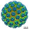

 PDBj
PDBj


 Assembly
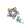


 Sample preparation
 Electron microscopy imaging
 Processing
