 Movie
Movie Controller

[English] 日本語
 Yorodumi
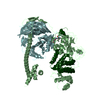
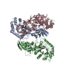
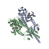
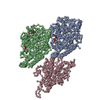
- PDB-3j6h: Nucleotide-free Kinesin motor domain complexed with GMPCPP-microtubule -
+ Open data

- Basic information
| Entry | Database: PDB / ID: 3j6h | ||||||
|---|---|---|---|---|---|---|---|
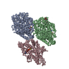
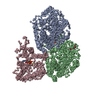
| Title | Nucleotide-free Kinesin motor domain complexed with GMPCPP-microtubule | ||||||
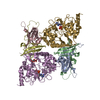
 Components Components |
| ||||||
 Keywords Keywords |  STRUCTURAL PROTEIN/MOTOR PROTEIN / Kinesin / Motor domain / Rigor-conformation / Nucleotide-free kinesin / Microtubule / GMPCPP-microtubule / tubulin / Axonal transport / STRUCTURAL PROTEIN-MOTOR PROTEIN complex STRUCTURAL PROTEIN/MOTOR PROTEIN / Kinesin / Motor domain / Rigor-conformation / Nucleotide-free kinesin / Microtubule / GMPCPP-microtubule / tubulin / Axonal transport / STRUCTURAL PROTEIN-MOTOR PROTEIN complex | ||||||
| Function / homology |  Function and homology information Function and homology informationdistal axon / anterograde dendritic transport of messenger ribonucleoprotein complex / anterograde dendritic transport of neurotransmitter receptor complex / anterograde axonal protein transport / intracellular mRNA localization / apolipoprotein receptor binding / plus-end-directed microtubule motor activity / motor neuron axon guidance / ciliary rootlet / postsynaptic cytosol ...distal axon / anterograde dendritic transport of messenger ribonucleoprotein complex / anterograde dendritic transport of neurotransmitter receptor complex / anterograde axonal protein transport / intracellular mRNA localization / apolipoprotein receptor binding / plus-end-directed microtubule motor activity / motor neuron axon guidance / ciliary rootlet / postsynaptic cytosol / kinesin complex / synaptic vesicle transport / microtubule motor activity / mRNA transport / axonal growth cone / axon cytoplasm / dendrite cytoplasm / axon guidance / Hydrolases; Acting on acid anhydrides; Acting on GTP to facilitate cellular and subcellular movement / Hydrolases; Acting on acid anhydrides; Acting on acid anhydrides to facilitate cellular and subcellular movement / structural constituent of cytoskeleton / microtubule cytoskeleton organization / mitotic cell cycle / microtubule binding / microtubule / hydrolase activity / neuron projection / GTPase activity / dendrite / neuronal cell body / GTP binding / ATP hydrolysis activity / ATP binding / metal ion binding / cytoplasmSimilarity search - Function | ||||||
| Biological species |  Mus musculus (house mouse)Sus scrofa (pig) Mus musculus (house mouse)Sus scrofa (pig) | ||||||
| Method | ELECTRON MICROSCOPY / single particle reconstruction / cryo EM / Resolution: 8.1 Å | ||||||
 Authors Authors | Morikawa, M. / Yajima, H. / Nitta, R. / Inoue, S. / Ogura, T. / Sato, C. / Hirokawa, N. | ||||||
 Citation Citation | Journal: EMBO J / Year: 2015 Title: X-ray and Cryo-EM structures reveal mutual conformational changes of Kinesin and GTP-state microtubules upon binding. Authors: Manatsu Morikawa / Hiroaki Yajima / Ryo Nitta / Shigeyuki Inoue / Toshihiko Ogura / Chikara Sato / Nobutaka Hirokawa /   Abstract: The molecular motor kinesin moves along microtubules using energy from ATP hydrolysis in an initial step coupled with ADP release. In neurons, kinesin-1/KIF5C preferentially binds to the GTP-state ...The molecular motor kinesin moves along microtubules using energy from ATP hydrolysis in an initial step coupled with ADP release. In neurons, kinesin-1/KIF5C preferentially binds to the GTP-state microtubules over GDP-state microtubules to selectively enter an axon among many processes; however, because the atomic structure of nucleotide-free KIF5C is unavailable, its molecular mechanism remains unresolved. Here, the crystal structure of nucleotide-free KIF5C and the cryo-electron microscopic structure of nucleotide-free KIF5C complexed with the GTP-state microtubule are presented. The structures illustrate mutual conformational changes induced by interaction between the GTP-state microtubule and KIF5C. KIF5C acquires the 'rigor conformation', where mobile switches I and II are stabilized through L11 and the initial portion of the neck-linker, facilitating effective ADP release and the weak-to-strong transition of KIF5C microtubule affinity. Conformational changes to tubulin strengthen the longitudinal contacts of the GTP-state microtubule in a similar manner to GDP-taxol microtubules. These results and functional analyses provide the molecular mechanism of the preferential binding of KIF5C to GTP-state microtubules. | ||||||
| History |
|
- Structure visualization
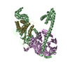
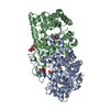
| Movie |
Movie viewer |
|---|---|
| Structure viewer | Molecule: MolmilJmol/JSmol |
- Downloads & links
-Download
| PDBx/mmCIF format | 3j6h.cif.gz | 215.5 KB | Display | PDBx/mmCIF format |
|---|---|---|---|---|
| PDB format | pdb3j6h.ent.gz | 156 KB | Display | PDB format |
| PDBx/mmJSON format | 3j6h.json.gz | Tree view | PDBx/mmJSON format | |
| Others |  Other downloads Other downloads |
-Validation report
| Arichive directory | https://data.pdbj.org/pub/pdb/validation_reports/j6/3j6hftp://data.pdbj.org/pub/pdb/validation_reports/j6/3j6h | HTTPS FTP |
|---|
-Related structure data
| Related structure data | 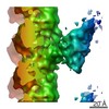 5916MC 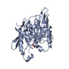 3wrdC 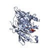 3x2tC M: map data used to model this data C: citing same article ( |
|---|---|
| Similar structure data |
-Links
 PDBj
PDBj


- Assembly
| Deposited unit | 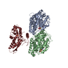
|
|---|---|
| 1 |
|
-Components
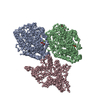
-Protein , 3 types, 3 molecules ABK
| #1: Protein | / Alpha-tubulin 1 / Tubulin alpha-1 chain Mass: 48436.625 Da / Num. of mol.: 1 / Fragment: UNP residues 2-437 / Source method: isolated from a natural source / Source: (natural) Sus scrofa (pig) / Tissue: brain / References: UniProt: P02550 |
|---|---|
| #2: Protein | Mass: 47809.746 Da / Num. of mol.: 1 / Fragment: UNP residues 2-427 / Source method: isolated from a natural source / Source: (natural) Sus scrofa (pig) / Tissue: brain / References: UniProt: P02554 |
| #3: Protein | Mass: 39615.137 Da / Num. of mol.: 1 / Fragment: UNP residues 1-345 Source method: isolated from a genetically manipulated source Source: (gene. exp.) Mus musculus (house mouse) / Gene: Kif5c / Plasmid: pET21b / Production host:  Escherichia coli (E. coli) / Strain (production host): BL21(DE3) / References: UniProt: P28738 Escherichia coli (E. coli) / Strain (production host): BL21(DE3) / References: UniProt: P28738 |
-Non-polymers , 4 types, 5 molecules 


| #4: Chemical |  Mass: 24.305 Da / Num. of mol.: 2 / Source method: obtained synthetically / Formula: Mg Mass: 24.305 Da / Num. of mol.: 2 / Source method: obtained synthetically / Formula: Mg#5: Chemical | ChemComp-GTP / | Guanosine triphosphate Mass: 523.180 Da / Num. of mol.: 1 / Source method: obtained synthetically / Formula: C10H16N5O14P3 / Comment: GTP, energy-carrying molecule*YM Mass: 523.180 Da / Num. of mol.: 1 / Source method: obtained synthetically / Formula: C10H16N5O14P3 / Comment: GTP, energy-carrying molecule*YM#6: Chemical | ChemComp-G2P / |  Mass: 521.208 Da / Num. of mol.: 1 / Source method: obtained synthetically / Formula: C11H18N5O13P3 / Comment: GMP-CPP, energy-carrying molecule analogue*YM Mass: 521.208 Da / Num. of mol.: 1 / Source method: obtained synthetically / Formula: C11H18N5O13P3 / Comment: GMP-CPP, energy-carrying molecule analogue*YM#7: Chemical | ChemComp-SO4 / | Sulfate Mass: 96.063 Da / Num. of mol.: 1 / Source method: obtained synthetically / Formula: SO4 Mass: 96.063 Da / Num. of mol.: 1 / Source method: obtained synthetically / Formula: SO4 |
|---|
-Details
| Sequence details | THIS SEQUENCE IS NATURAL VARIANT. |
|---|
-Experimental details
-Experiment
| Experiment | Method: ELECTRON MICROSCOPY |
|---|---|
| EM experiment | Aggregation state: PARTICLE / 3D reconstruction method: single particle reconstruction |
- Sample preparation
| Component | Name: Kinesin motor domain complexed with GMPCPP-microtubule Type: COMPLEX |
|---|---|
| Buffer solution | pH: 6.8 |
| Specimen | Embedding applied: NO / Shadowing applied: NO / Staining applied: NO / Vitrification applied: YES |
| Vitrification | Instrument: LEICA KF80 / Cryogen name: ETHANE |
- Electron microscopy imaging
| Microscopy | Model: JEOL 2010F / Date: Jan 1, 2012 |
|---|---|
| Electron gun | Electron source: FIELD EMISSION GUN / Accelerating voltage: 200 kV / Illumination mode: FLOOD BEAM |
| Electron lens | Mode: BRIGHT FIELDBright-field microscopy / Nominal magnification: 40000 X / Calibrated magnification: 40000 X / Nominal defocus max: 2600 nm / Nominal defocus min: 1200 nm / Cs: 3.3 mm |
| Specimen holder | Temperature: 100 K / Tilt angle max: 0 ° / Tilt angle min: 0 ° |
| Image recording | Electron dose: 10 e/Å2 / Film or detector model: KODAK SO-163 FILM |
| Radiation | Protocol: SINGLE WAVELENGTH / Monochromatic (M) / Laue (L): M / Scattering type: x-ray |
| Radiation wavelength | Relative weight: 1 |
- Processing
| EM software |
| ||||||||||||||||||||||||||||
|---|---|---|---|---|---|---|---|---|---|---|---|---|---|---|---|---|---|---|---|---|---|---|---|---|---|---|---|---|---|
| CTF correction | Details: Each filament | ||||||||||||||||||||||||||||
| Symmetry | Point symmetry: C1 (asymmetric) | ||||||||||||||||||||||||||||
| 3D reconstruction | Method: Single ParticleSingle particle analysis / Resolution: 8.1 Å / Num. of particles: 302000 / Nominal pixel size: 2.5 Å / Actual pixel size: 2.5 Å / Num. of class averages: 18 / Symmetry type: POINT | ||||||||||||||||||||||||||||
| Atomic model building | Protocol: RIGID BODY FIT / Space: REAL Target criteria: Cross-correlation, Average map value, Atoms inside the contour Details: METHOD--Local refinement, Domain fitting REFINEMENT PROTOCOL--Rigid body refinement DETAILS--Initial local fitting was done using Chimera and for some loops Modeller was used. | ||||||||||||||||||||||||||||
| Atomic model building |
| ||||||||||||||||||||||||||||
| Refinement step | Cycle: LAST
|

