Movie
Movie Controller

[English] 日本語
 Yorodumi
- PDB-3j3p: Conformational Shift of a Major Poliovirus Antigen Confirmed by I... -
+ Open data

- Basic information
| Entry | Database: PDB / ID: 3j3p | ||||||
|---|---|---|---|---|---|---|---|
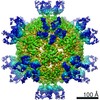
| Title | Conformational Shift of a Major Poliovirus Antigen Confirmed by Immuno-Cryogenic Electron Microscopy: 135S Poliovirus and C3-Fab Complex | ||||||
 Components Components |
| ||||||
 Keywords Keywords | VIRUS/IMMUNE SYSTEM / 135S cell-entry intermediate particle /  antibody-antigen interaction / antibody-protein interaction / picornavirus / virus-antibody interaction / antibody neutralization / neutralizing antibody interaction / conformational change / VIRUS-IMMUNE SYSTEM complex antibody-antigen interaction / antibody-protein interaction / picornavirus / virus-antibody interaction / antibody neutralization / neutralizing antibody interaction / conformational change / VIRUS-IMMUNE SYSTEM complex | ||||||
| Function / homology |  Function and homology information Function and homology informationsymbiont-mediated suppression of host translation initiation / positive regulation of B cell activation / early endosome to late endosome transport / humoral immune response mediated by circulating immunoglobulin / phagocytosis, recognition / positive regulation of type IIa hypersensitivity / regulation of proteolysis / positive regulation of type I hypersensitivity / antibody-dependent cellular cytotoxicity / Fc-gamma receptor I complex binding ...symbiont-mediated suppression of host translation initiation / positive regulation of B cell activation / early endosome to late endosome transport / humoral immune response mediated by circulating immunoglobulin / phagocytosis, recognition / positive regulation of type IIa hypersensitivity / regulation of proteolysis / positive regulation of type I hypersensitivity / antibody-dependent cellular cytotoxicity / Fc-gamma receptor I complex binding / phagocytosis, engulfment / endosome to lysosome transport / positive regulation of endocytosis / immunoglobulin complex, circulating / IgG immunoglobulin complex / immunoglobulin receptor binding / symbiont-mediated suppression of host cytoplasmic pattern recognition receptor signaling pathway via inhibition of MDA-5 activity / symbiont-mediated suppression of host cytoplasmic pattern recognition receptor signaling pathway via inhibition of RIG-I activity / immunoglobulin mediated immune response / antigen processing and presentation / positive regulation of phagocytosis / complement activation, classical pathway / picornain 2A / symbiont-mediated suppression of host mRNA export from nucleus / symbiont-mediated suppression of host cytoplasmic pattern recognition receptor signaling pathway via inhibition of MAVS activity / antigen binding / symbiont genome entry into host cell via pore formation in plasma membrane / picornain 3C / multivesicular body / ribonucleoside triphosphate phosphatase activity / T=pseudo3 icosahedral viral capsid / host cell cytoplasmic vesicle membrane / response to bacterium / endocytosis involved in viral entry into host cell / : / positive regulation of immune response / nucleoside-triphosphate phosphatase / protein complex oligomerization / monoatomic ion channel activity / antibacterial humoral response / RNA helicase activity / induction by virus of host autophagy / RNA-directed RNA polymerase / symbiont-mediated suppression of host gene expression / viral RNA genome replication / cysteine-type endopeptidase activity / RNA-dependent RNA polymerase activity / DNA-templated transcription / host cell nucleus / structural molecule activity / virion attachment to host cell / proteolysis / extracellular space / RNA binding / ATP binding / membrane / metal ion binding / plasma membrane / cytosolSimilarity search - Function | ||||||
| Biological species |  Mus musculus (house mouse) Mus musculus (house mouse) Human poliovirus 1 Human poliovirus 1 | ||||||
| Method | ELECTRON MICROSCOPY / single particle reconstruction / cryo EM / Resolution: 9.1 Å | ||||||
 Authors Authors | Lin, J. / Cheng, N. / Hogle, J.M. / Steven, A.C. / Belnap, D.M. | ||||||
 Citation Citation | Journal: J Immunol / Year: 2013 Title: Conformational shift of a major poliovirus antigen confirmed by immuno-cryogenic electron microscopy. Authors: Jun Lin / Naiqian Cheng / James M Hogle / Alasdair C Steven / David M Belnap /  Abstract: Small, interfacial conformational changes occur in some Ag-Ab interactions. Using cryogenic electron microscopy (cryo-EM), we have demonstrated such changes in a major antigenic site of a poliovirus ...Small, interfacial conformational changes occur in some Ag-Ab interactions. Using cryogenic electron microscopy (cryo-EM), we have demonstrated such changes in a major antigenic site of a poliovirus capsid protein. During cell entry, native human poliovirus (160S particle) converts to a cell entry intermediate (135S particle) and later to an RNA-released (80S) particle. By mixing particles with Fabs of the neutralizing C3 mAb, we labeled the external loop connecting the B and C β-strands (BC loop) of the capsid protein VP1 (residues 95-105) in the 160S and 135S states. We then determined three-dimensional structures by cryo-EM and enhanced their interpretability by fitting high-resolution coordinates of C3 Fab and the capsid proteins into the density maps. Binding of C3 to either 160S or 135S particles caused residues of the BC loop, located on the tip of a prominent peak known as the "mesa," to move by an estimated 5 Å. C3 Abs are neutralizing and can bind bivalently. The orientation of the bound Fabs in our reconstructions suggests that C3 neutralizes poliovirus by binding two adjacent BC loops on the same mesa and inhibiting conformational changes in the viral capsid. | ||||||
| History |
| ||||||
| Remark 650 | HELIX DETERMINATION METHOD: AUTHOR DETERMINED | ||||||
| Remark 700 | SHEET DETERMINATION METHOD: AUTHOR DETERMINED |
- Structure visualization
| Movie |
Movie viewer |
|---|---|
| Structure viewer | Molecule: MolmilJmol/JSmol |
- Downloads & links
-Download
| PDBx/mmCIF format | 3j3p.cif.gz | 57.2 KB | Display | PDBx/mmCIF format |
|---|---|---|---|---|
| PDB format | pdb3j3p.ent.gz | 28.8 KB | Display | PDB format |
| PDBx/mmJSON format | 3j3p.json.gz | Tree view | PDBx/mmJSON format | |
| Others |  Other downloads Other downloads |
-Validation report
| Arichive directory | https://data.pdbj.org/pub/pdb/validation_reports/j3/3j3pftp://data.pdbj.org/pub/pdb/validation_reports/j3/3j3p | HTTPS FTP |
|---|
-Related structure data
| Related structure data |  5292MC 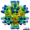 5291C  5293C  3j3oC M: map data used to model this data C: citing same article ( |
|---|---|
| Similar structure data |
-Links
 PDBj
PDBj


- Assembly
| Deposited unit | 
|
|---|---|
| 1 | x 60
|
| 2 |
|
| 3 | x 5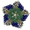
|
| 4 | x 6
|
| 5 | 
|
| Symmetry | Point symmetry: (Schoenflies symbol: I (icosahedral)) |
-Components
| #1: Antibody | Mass: 24080.750 Da / Num. of mol.: 1 / Fragment: Fab / Source method: isolated from a natural source / Source: (natural) Mus musculus (house mouse) |
|---|---|
| #2: Antibody | Mass: 23478.137 Da / Num. of mol.: 1 / Fragment: Fab / Source method: isolated from a natural source / Source: (natural) Mus musculus (house mouse) / References: UniProt: P01865*PLUS |
| #3: Protein | Mass: 33488.613 Da / Num. of mol.: 1 / Fragment: UNP residues 580-881 / Source method: isolated from a natural source / Source: (natural) Human poliovirus 1 / Strain: Mahoney / References: UniProt: P03300 |
| #4: Protein | Mass: 30075.783 Da / Num. of mol.: 1 / Fragment: UNP residues 70-341 / Source method: isolated from a natural source / Source: (natural) Human poliovirus 1 / Strain: Mahoney / References: UniProt: P03300 |
| #5: Protein | Mass: 26547.482 Da / Num. of mol.: 1 / Fragment: UNP residues 342-579 / Source method: isolated from a natural source / Source: (natural) Human poliovirus 1 / Strain: Mahoney / References: UniProt: P03300 |
-Experimental details
-Experiment
| Experiment | Method: ELECTRON MICROSCOPY |
|---|---|
| EM experiment | Aggregation state: PARTICLE / 3D reconstruction method: single particle reconstruction |
- Sample preparation
| Component | Name: Poliovirus 135S particle and C3 Fab complex / Type: VIRUS / Details: 135S icosahedral particle with Fab |
|---|---|
| Details of virus | Empty: NO / Enveloped: NO / Host category: VERTEBRATES / Isolate: STRAIN / Type: VIRION |
| Natural host | Organism: Homo sapiens |
| Buffer solution | Name: 20 mM Tris, 2 mM CaCl2 / pH: 7.5 / Details: 20 mM Tris, 2 mM CaCl2 |
| Specimen | Embedding applied: NO / Shadowing applied: NO / Staining applied: NO / Vitrification applied: YES |
| Vitrification | Cryogen name: ETHANE Details: Vitrification carried out in ambient atmosphere. Ethane cooled by liquid nitrogen. Method: Blotted manually before plunging |
- Electron microscopy imaging
| Microscopy | Model: FEI/PHILIPS CM200FEG |
|---|---|
| Electron gun | Electron source: FIELD EMISSION GUN / Accelerating voltage: 120 kV / Illumination mode: FLOOD BEAM |
| Electron lens | Mode: BRIGHT FIELDBright-field microscopy / Nominal magnification: 38000 X / Calibrated magnification: 38000 X / Nominal defocus max: 1630 nm / Nominal defocus min: 1120 nm / Cs: 2 mm / Astigmatism: Bsoft / Camera length: 0 mm |
| Specimen holder | Specimen holder model: GATAN LIQUID NITROGEN Specimen holder type: Side entry liquid nitrogen-cooled cryo specimen holder |
| Image recording | Electron dose: 14 e/Å2 / Film or detector model: KODAK SO-163 FILM |
| Image scans | Num. digital images: 4 |
| Radiation | Protocol: SINGLE WAVELENGTH / Monochromatic (M) / Laue (L): M / Scattering type: x-ray |
| Radiation wavelength | Relative weight: 1 |
- Processing
| EM software |
| ||||||||||||||||||||||||||||||
|---|---|---|---|---|---|---|---|---|---|---|---|---|---|---|---|---|---|---|---|---|---|---|---|---|---|---|---|---|---|---|---|
| CTF correction | Details: CTF and decay correction of each particle | ||||||||||||||||||||||||||||||
| Symmetry | Point symmetry: I (icosahedral) | ||||||||||||||||||||||||||||||
| 3D reconstruction | Method: Fourier Bessel / Resolution: 9.1 Å / Resolution method: FSC 0.5 CUT-OFF / Num. of particles: 9810 / Nominal pixel size: 1.84 Å / Actual pixel size: 1.84 Å Details: Reconstruction computed from focal pairs. Pairs not summed for reconstruction calculation. Symmetry type: POINT | ||||||||||||||||||||||||||||||
| Atomic model building |
| ||||||||||||||||||||||||||||||
| Atomic model building | Source name: PDB / Type: experimental model
| ||||||||||||||||||||||||||||||
| Refinement step | Cycle: LAST
|