 Movie
Movie Controller


+ Open data

- Basic information
| Entry | Database: PDB / ID: 3j2q | |||||||||
|---|---|---|---|---|---|---|---|---|---|---|
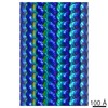
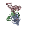
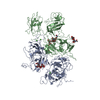
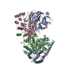
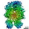
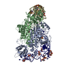
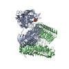
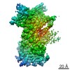
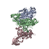
| Title | Model of membrane-bound factor VIII organized in 2D crystals | |||||||||
 Components Components | (Coagulation factor VIII ...) x 2 | |||||||||
 Keywords Keywords | BLOOD CLOTTING / blood coagulation / cofactor / factor VIII / hemophilia | |||||||||
| Function / homology |  Function and homology information Function and homology informationDefective F8 accelerates dissociation of the A2 domain / Defective F8 binding to the cell membrane / Defective F8 secretion / Defective F8 sulfation at Y1699 / Gamma carboxylation, hypusinylation, hydroxylation, and arylsulfatase activation / Defective F8 binding to von Willebrand factor / blood coagulation, intrinsic pathway / Cargo concentration in the ER / Defective factor IX causes thrombophilia / Defective cofactor function of FVIIIa variant ...Defective F8 accelerates dissociation of the A2 domain / Defective F8 binding to the cell membrane / Defective F8 secretion / Defective F8 sulfation at Y1699 / Gamma carboxylation, hypusinylation, hydroxylation, and arylsulfatase activation / Defective F8 binding to von Willebrand factor / blood coagulation, intrinsic pathway / Cargo concentration in the ER / Defective factor IX causes thrombophilia / Defective cofactor function of FVIIIa variant / Defective F9 variant does not activate FX / COPII-mediated vesicle transport / COPII-coated ER to Golgi transport vesicle / Defective F8 cleavage by thrombin / Common Pathway of Fibrin Clot Formation / Intrinsic Pathway of Fibrin Clot Formation / endoplasmic reticulum-Golgi intermediate compartment membrane / platelet alpha granule lumen / acute-phase response / Golgi lumen / blood coagulation / Platelet degranulation / oxidoreductase activity / copper ion binding / endoplasmic reticulum lumen / extracellular space / extracellular region / plasma membrane Similarity search - Function | |||||||||
| Biological species |  Homo sapiens (human) Homo sapiens (human) | |||||||||
| Method | ELECTRON CRYSTALLOGRAPHY / electron crystallography / cryo EM / Resolution: 15 Å | |||||||||
 Authors Authors | Stoilova-Mcphie, S. / Lynch, G.C. / Ludtke, S. / Pettitt, B.M. | |||||||||
 Citation Citation | Journal: Biopolymers / Year: 2013 Title: Domain organization of membrane-bound factor VIII. Authors: Svetla Stoilova-McPhie / Gillian C Lynch / Steven Ludtke / B Montgomery Pettitt /  Abstract: Factor VIII (FVIII) is the blood coagulation protein which when defective or deficient causes for hemophilia A, a severe hereditary bleeding disorder. Activated FVIII (FVIIIa) is the cofactor to the ...Factor VIII (FVIII) is the blood coagulation protein which when defective or deficient causes for hemophilia A, a severe hereditary bleeding disorder. Activated FVIII (FVIIIa) is the cofactor to the serine protease factor IXa (FIXa) within the membrane-bound Tenase complex, responsible for amplifying its proteolytic activity more than 100,000 times, necessary for normal clot formation. FVIII is composed of two noncovalently linked peptide chains: a light chain (LC) holding the membrane interaction sites and a heavy chain (HC) holding the main FIXa interaction sites. The interplay between the light and heavy chains (HCs) in the membrane-bound state is critical for the biological efficiency of FVIII. Here, we present our cryo-electron microscopy (EM) and structure analysis studies of human FVIII-LC, when helically assembled onto negatively charged single lipid bilayer nanotubes. The resolved FVIII-LC membrane-bound structure supports aspects of our previously proposed FVIII structure from membrane-bound two-dimensional (2D) crystals, such as only the C2 domain interacts directly with the membrane. The LC is oriented differently in the FVIII membrane-bound helical and 2D crystal structures based on EM data, and the existing X-ray structures. This flexibility of the FVIII-LC domain organization in different states is discussed in the light of the FVIIIa-FIXa complex assembly and function. #1: Journal: Blood / Year: 2002 Title: 3-Dimensional structure of membrane-bound coagulation factor VIII: modeling of the factor VIII heterodimer within a 3-dimensional density map derived by electron crystallography. Authors: Svetla Stoilova-McPhie / Bruno O Villoutreix / Koen Mertens / Geoffrey Kemball-Cook / Andreas Holzenburg / Abstract: Despite recent studies, the organization of coagulation factor VIII (FVIII) on a phospholipid (PL) membrane is not known in detail. Thus, 2-dimensional (2D) crystals of human FVIII lacking the B ...Despite recent studies, the organization of coagulation factor VIII (FVIII) on a phospholipid (PL) membrane is not known in detail. Thus, 2-dimensional (2D) crystals of human FVIII lacking the B domain were prepared for electron microscopy onto negatively charged PL monolayers. The 3-dimensional (3D) density map of the PL-bound FVIII protein was calculated at 1.5 nm. Existing atomic data and models for FVIII domains were fitted unambiguously within the 3D density map of the molecule. FVIII domains arrangement followed a compact spiral organization with the A3 domains in close association with the C1 and C2 domains near the PL surface. Viewed toward the membrane the A domains' heterotrimer is oriented side-on with the pseudo-3-fold axis almost parallel to the PL surface and A1 fully covering C1. The C2 domain is partially overlapped by the A2 domain of an adjacent molecule in the 2D crystal, favoring close packing. Viewed parallel to the membrane, C2 is slightly inclined to the PL surface covering an area of 12 nm(2). Four C2 loops are embedded within the lipid monolayer at about 0.7 to 1.0 nm depth. C1 forms almost a right angle with C2, its long axis nearly parallel to the membrane. The proposed structure for membrane-bound FVIII results from modeling of the FVIII domains within a 3D density map obtained from electron crystallography and accords with the main biochemical and structural information known to date. A model is proposed for FVIIIa and factor IXa assembly within the membrane-bound factor X-activating complex. (Blood. 2002;99:1215-1223) | |||||||||
| History |
|
- Structure visualization
| Movie |
Movie viewer |
|---|---|
| Structure viewer | Molecule: MolmilJmol/JSmol |
- Downloads & links
-Download
| PDBx/mmCIF format | 3j2q.cif.gz | 277.5 KB | Display | PDBx/mmCIF format |
|---|---|---|---|---|
| PDB format | pdb3j2q.ent.gz | 185.7 KB | Display | PDB format |
| PDBx/mmJSON format | 3j2q.json.gz | Tree view | PDBx/mmJSON format | |
| Others |  Other downloads Other downloads |
-Validation report
| Summary document | 3j2q_validation.pdf.gz | 596.2 KB | Display | wwPDB validaton report |
|---|---|---|---|---|
| Full document | 3j2q_full_validation.pdf.gz | 940.5 KB | Display | |
| Data in XML | 3j2q_validation.xml.gz | 86.8 KB | Display | |
| Data in CIF | 3j2q_validation.cif.gz | 116.4 KB | Display | |
| Arichive directory | https://data.pdbj.org/pub/pdb/validation_reports/j2/3j2qftp://data.pdbj.org/pub/pdb/validation_reports/j2/3j2q | HTTPS FTP |
-Related structure data


-Links
 PDBj
PDBj


- Assembly
| Deposited unit | 
| ||||||||
|---|---|---|---|---|---|---|---|---|---|
| 1 |
| ||||||||
| Unit cell |
|
-Components
-Coagulation factor VIII ... , 2 types, 2 molecules AB
| #1: Protein | Mass: 86455.742 Da / Num. of mol.: 1 / Fragment: UNP residues 20-764 Source method: isolated from a genetically manipulated source Source: (gene. exp.) Homo sapiens (human) / Gene: F8, F8C / Cell (production host): ovary / Production host:   Cricetulus griseus (Chinese hamster) / References: UniProt: P00451 Cricetulus griseus (Chinese hamster) / References: UniProt: P00451 |
|---|---|
| #2: Protein | Mass: 79050.594 Da / Num. of mol.: 1 / Fragment: UNP residues 1668-2351 Source method: isolated from a genetically manipulated source Source: (gene. exp.) Homo sapiens (human) / Gene: F8, F8C / Cell (production host): ovary / Production host: Cricetulus griseus (Chinese hamster) / References: UniProt: P00451 |
-Sugars , 3 types, 4 molecules 
| #3: Polysaccharide | 2-acetamido-2-deoxy-beta-D-glucopyranose-(1-4)-2-acetamido-2-deoxy-beta-D-glucopyranose Source method: isolated from a genetically manipulated source |
|---|---|
| #4: Polysaccharide | alpha-D-mannopyranose-(1-3)-[beta-D-mannopyranose-(1-6)]beta-D-mannopyranose-(1-4)-2-acetamido-2- ...alpha-D-mannopyranose-(1-3)-[beta-D-mannopyranose-(1-6)]beta-D-mannopyranose-(1-4)-2-acetamido-2-deoxy-beta-D-glucopyranose Source method: isolated from a genetically manipulated source |
| #7: Sugar |  Type: D-saccharide, beta linking / Mass: 221.208 Da / Num. of mol.: 2 Type: D-saccharide, beta linking / Mass: 221.208 Da / Num. of mol.: 2Source method: isolated from a genetically manipulated source Formula: C8H15NO6 |
-Non-polymers , 2 types, 3 molecules 

| #5: Chemical |  Mass: 63.546 Da / Num. of mol.: 2 / Source method: obtained synthetically / Formula: Cu Mass: 63.546 Da / Num. of mol.: 2 / Source method: obtained synthetically / Formula: Cu#6: Chemical | ChemComp-CA / |  Mass: 40.078 Da / Num. of mol.: 1 / Source method: obtained synthetically / Formula: Ca Mass: 40.078 Da / Num. of mol.: 1 / Source method: obtained synthetically / Formula: Ca |
|---|
-Details
| Sequence details | THE AUTHORS STATE THAT F1899L IS A NATURAL VARIANT. |
|---|
-Experimental details
-Experiment
| Experiment | Method: ELECTRON CRYSTALLOGRAPHY |
|---|---|
| EM experiment | Aggregation state: 2D ARRAY / 3D reconstruction method: electron crystallography |
- Sample preparation
| Component | Name: membrane-bound factor VIII / Type: COMPLEX |
|---|---|
| Specimen | Embedding applied: NO / Shadowing applied: NO / Staining applied: NO / Vitrification applied: YES |
-Data collection
| Microscopy | Model: JEOL 2010F |
|---|---|
| Electron gun | Electron source:  FIELD EMISSION GUN / Accelerating voltage: 200 kV / Illumination mode: FLOOD BEAM FIELD EMISSION GUN / Accelerating voltage: 200 kV / Illumination mode: FLOOD BEAM |
| Electron lens | Mode: BRIGHT FIELD / Nominal magnification: 52000 X |
| Image recording | Electron dose: 16 e/Å2 |
| Radiation | Protocol: SINGLE WAVELENGTH / Monochromatic (M) / Laue (L): M / Scattering type: electron |
| Radiation wavelength | Relative weight: 1 |
- Processing
| 3D reconstruction | Resolution: 15 Å / Symmetry type: 2D CRYSTAL | ||||||||||||
|---|---|---|---|---|---|---|---|---|---|---|---|---|---|
| Refinement step | Cycle: LAST
|
