 Movie
Movie Controller

[English] 日本語
 Yorodumi
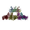
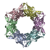
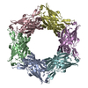
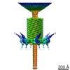
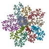
- PDB-3j2n: The X-ray structure of the gp15 hexamer and the model of the gp18... -
+ Open data

- Basic information
| Entry | Database: PDB / ID: 3j2n | ||||||
|---|---|---|---|---|---|---|---|
| Title | The X-ray structure of the gp15 hexamer and the model of the gp18 protein fitted into the cryo-EM reconstruction of the contracted T4 tail | ||||||
 Components Components |
| ||||||
 Keywords Keywords | VIRAL PROTEIN / bacteriophage T4 / phage tail terminator protein / phage sheath protein | ||||||
| Function / homology |  Function and homology information Function and homology informationvirus tail, sheath / symbiont genome ejection through host cell envelope, contractile tail mechanism / virion component Similarity search - Function | ||||||
| Biological species |  Enterobacteria phage T4 (virus) Enterobacteria phage T4 (virus) | ||||||
| Method | ELECTRON MICROSCOPY / single particle reconstruction / cryo EM / Resolution: 16 Å | ||||||
 Authors Authors | Fokine, A. / Zhang, Z. / Kanamaru, S. / Bowman, V.D. / Aksyuk, A. / Arisaka, F. / Rao, V.B. / Rossmann, M.G. | ||||||
 Citation Citation | Journal: J Mol Biol / Year: 2013 Title: The molecular architecture of the bacteriophage T4 neck. Authors: Andrei Fokine / Zhihong Zhang / Shuji Kanamaru / Valorie D Bowman / Anastasia A Aksyuk / Fumio Arisaka / Venigalla B Rao / Michael G Rossmann /  Abstract: A hexamer of the bacteriophage T4 tail terminator protein, gp15, attaches to the top of the phage tail stabilizing the contractile sheath and forming the interface for binding of the independently ...A hexamer of the bacteriophage T4 tail terminator protein, gp15, attaches to the top of the phage tail stabilizing the contractile sheath and forming the interface for binding of the independently assembled head. Here we report the crystal structure of the gp15 hexamer, describe its interactions in T4 virions that have either an extended tail or a contracted tail, and discuss its structural relationship to other phage proteins. The neck of T4 virions is decorated by the "collar" and "whiskers", made of fibritin molecules. Fibritin acts as a chaperone helping to attach the long tail fibers to the virus during the assembly process. The collar and whiskers are environment-sensing devices, regulating the retraction of the long tail fibers under unfavorable conditions, thus preventing infection. Cryo-electron microscopy analysis suggests that twelve fibritin molecules attach to the phage neck with six molecules forming the collar and six molecules forming the whiskers. #1: Journal: Cell / Year: 2004Title: Three-dimensional rearrangement of proteins in the tail of bacteriophage T4 on infection of its host. Authors: Petr G Leiman / Paul R Chipman / Victor A Kostyuchenko / Vadim V Mesyanzhinov / Michael G Rossmann / Abstract: The contractile tail of bacteriophage T4 undergoes major structural transitions when the virus attaches to the host cell surface. The baseplate at the distal end of the tail changes from a hexagonal ...The contractile tail of bacteriophage T4 undergoes major structural transitions when the virus attaches to the host cell surface. The baseplate at the distal end of the tail changes from a hexagonal to a star shape. This causes the sheath around the tail tube to contract and the tail tube to protrude from the baseplate and pierce the outer cell membrane and the cell wall before reaching the inner cell membrane for subsequent viral DNA injection. Analogously, the T4 tail can be contracted by treatment with 3 M urea. The structure of the T4 contracted tail, including the head-tail joining region, has been determined by cryo-electron microscopy to 17 A resolution. This 1200 A-long, 20 MDa structure has been interpreted in terms of multiple copies of its approximately 20 component proteins. A comparison with the metastable hexagonal baseplate of the mature virus shows that the baseplate proteins move as rigid bodies relative to each other during the structural change. | ||||||
| History |
|
- Structure visualization
| Movie |
Movie viewer |
|---|---|
| Structure viewer | Molecule: MolmilJmol/JSmol |
- Downloads & links
-Download
| PDBx/mmCIF format | 3j2n.cif.gz | 915 KB | Display | PDBx/mmCIF format |
|---|---|---|---|---|
| PDB format | pdb3j2n.ent.gz | 760.4 KB | Display | PDB format |
| PDBx/mmJSON format | 3j2n.json.gz | Tree view | PDBx/mmJSON format | |
| Others |  Other downloads Other downloads |
-Validation report
| Summary document | 3j2n_validation.pdf.gz | 1010.5 KB | Display | wwPDB validaton report |
|---|---|---|---|---|
| Full document | 3j2n_full_validation.pdf.gz | 1.3 MB | Display | |
| Data in XML | 3j2n_validation.xml.gz | 164.6 KB | Display | |
| Data in CIF | 3j2n_validation.cif.gz | 244.4 KB | Display | |
| Arichive directory | https://data.pdbj.org/pub/pdb/validation_reports/j2/3j2nftp://data.pdbj.org/pub/pdb/validation_reports/j2/3j2n | HTTPS FTP |
-Related structure data


-Links
 PDBj
PDBj- Assembly
| Deposited unit | 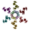
|
|---|---|
| 1 |
|
-Components
| #1: Protein | Mass: 31587.486 Da / Num. of mol.: 6 Source method: isolated from a genetically manipulated source Source: (gene. exp.) Enterobacteria phage T4 (virus) / Gene: 15 / Production host:  #2: Protein | Mass: 71289.484 Da / Num. of mol.: 6 / Mutation: R510P Source method: isolated from a genetically manipulated source Source: (gene. exp.) Enterobacteria phage T4 (virus) / Gene: 18 / Production host: Sequence details | THE AUTHORS STATE THAT D100E, G148A, N150I, Y151I, E301G, A399V, AND H454Y ARE NATURAL VARIANTS AS ...THE AUTHORS STATE THAT D100E, G148A, N150I, Y151I, E301G, A399V, AND H454Y ARE NATURAL VARIANTS AS PER PDB ENTRY 3FOA. | |
|---|
-Experimental details
-Experiment
| Experiment | Method: ELECTRON MICROSCOPY |
|---|---|
| EM experiment | Aggregation state: PARTICLE / 3D reconstruction method: single particle reconstruction |
- Sample preparation
| Component |
| ||||||||||||||||||||
|---|---|---|---|---|---|---|---|---|---|---|---|---|---|---|---|---|---|---|---|---|---|
| Buffer solution | Name: 3 M urea / pH: 7.5 / Details: 3 M urea | ||||||||||||||||||||
| Specimen | Conc.: 5 mg/ml / Embedding applied: NO / Shadowing applied: NO / Staining applied: NO / Vitrification applied: YES | ||||||||||||||||||||
| Specimen support | Details: 200 mesh copper grid | ||||||||||||||||||||
| Vitrification | Cryogen name: ETHANE / Temp: 100 K |
- Electron microscopy imaging
| Microscopy | Model: FEI/PHILIPS CM300FEG/T / Date: Jan 6, 2002 |
|---|---|
| Electron gun | Electron source:  FIELD EMISSION GUN / Accelerating voltage: 300 kV / Illumination mode: SPOT SCAN FIELD EMISSION GUN / Accelerating voltage: 300 kV / Illumination mode: SPOT SCAN |
| Electron lens | Mode: BRIGHT FIELD / Nominal magnification: 45000 X / Calibrated magnification: 47000 X / Nominal defocus max: 3400 nm / Nominal defocus min: 500 nm |
| Specimen holder | Specimen holder model: GATAN LIQUID NITROGEN Specimen holder type: Side entry liquid nitrogen-cooled cryo specimen holder Temperature: 100 K / Tilt angle max: 0 ° / Tilt angle min: 0 ° |
| Image recording | Electron dose: 20 e/Å2 / Film or detector model: KODAK SO-163 FILM / Details: Kodak SO163 film |
| EM imaging optics | Energyfilter name: FEI |
| Image scans | Num. digital images: 100 |
| Radiation | Protocol: SINGLE WAVELENGTH / Monochromatic (M) / Laue (L): M / Scattering type: x-ray |
| Radiation wavelength | Relative weight: 1 |
- Processing
| EM software |
| ||||||||||||
|---|---|---|---|---|---|---|---|---|---|---|---|---|---|
| CTF correction | Details: Each particle | ||||||||||||
| Symmetry | Point symmetry: C6 (6 fold cyclic) | ||||||||||||
| 3D reconstruction | Resolution: 16 Å / Resolution method: FSC 0.5 CUT-OFF / Num. of particles: 1965 / Nominal pixel size: 3.93 Å / Actual pixel size: 3.93 Å / Symmetry type: POINT | ||||||||||||
| Atomic model building | Space: REAL | ||||||||||||
| Refinement step | Cycle: LAST
|
