 Movie
Movie Controller


+ Open data

- Basic information
| Entry | Database: PDB / ID: 3ix0 | ||||||
|---|---|---|---|---|---|---|---|
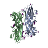
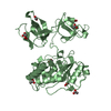
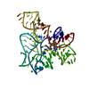
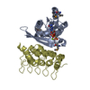
| Title | Crystal structure of human seminal plasma protein PSP94 | ||||||
 Components Components | Beta-microseminoprotein | ||||||
 Keywords Keywords |  ANTITUMOR PROTEIN / APOPTOSIS / beta sheet / Greek key motif / Disulfide bond / Secreted / PROTEIN BINDING ANTITUMOR PROTEIN / APOPTOSIS / beta sheet / Greek key motif / Disulfide bond / Secreted / PROTEIN BINDING | ||||||
| Function / homology |  Function and homology information Function and homology information | ||||||
| Biological species |  Homo sapiens (human) Homo sapiens (human) | ||||||
| Method | X-RAY DIFFRACTION / SYNCHROTRON / SIRAS / Resolution: 2.3 Å | ||||||
 Authors Authors | Kumar, M. / Kumar, A. / Jagtap, D.D. / Mahale, S.D. | ||||||
 Citation Citation | Journal: J.Mol.Biol. / Year: 2010 Title: Crystal structure of prostate secretory protein PSP94 shows an edge-to-edge association of two monomers to form a homodimer Authors: Kumar, A. / Jagtap, D.D. / Mahale, S.D. / Kumar, M. #1: Journal: Acta Crystallogr.,Sect.F / Year: 2009 Title: Crystallization and preliminary X-ray diffraction analysis of human seminal plasma protein PSP94 Authors: Kumar, M. / Jagtap, D.D. / Mahale, S.D. / Prashar, V. / Kumar, A. / Das, A. / Bihani, S.C. / Ferrer, J.L. / Hosur, M.V. / Ramanadham, M. | ||||||
| History |
|
- Structure visualization
| Structure viewer | Molecule: MolmilJmol/JSmol |
|---|
- Downloads & links
-Download
| PDBx/mmCIF format | 3ix0.cif.gz | 157.2 KB | Display | PDBx/mmCIF format |
|---|---|---|---|---|
| PDB format | pdb3ix0.ent.gz | 132.6 KB | Display | PDB format |
| PDBx/mmJSON format | 3ix0.json.gz | Tree view | PDBx/mmJSON format | |
| Others |  Other downloads Other downloads |
-Validation report
| Arichive directory | https://data.pdbj.org/pub/pdb/validation_reports/ix/3ix0ftp://data.pdbj.org/pub/pdb/validation_reports/ix/3ix0 | HTTPS FTP |
|---|
-Related structure data
| Similar structure data |
|---|


-Links
 PDBj
PDBj- Assembly
| Deposited unit | 
| ||||||||
|---|---|---|---|---|---|---|---|---|---|
| 1 | 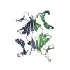
| ||||||||
| 2 | 
| ||||||||
| Unit cell |
| ||||||||
| Details | Either (Chain A and B) OR (chain C and D) make biological unit |
-Components
| #1: Protein | Mass: 10784.274 Da / Num. of mol.: 4 / Source method: isolated from a natural source / Source: (natural) Homo sapiens (human) / Tissue: Human Seminal Plasma / References: UniProt: P08118#2: Water | ChemComp-HOH / | Water Mass: 18.015 Da / Num. of mol.: 241 / Source method: isolated from a natural source / Formula: H2O Mass: 18.015 Da / Num. of mol.: 241 / Source method: isolated from a natural source / Formula: H2O |
|---|
-Experimental details
-Experiment
| Experiment | Method: X-RAY DIFFRACTION / Number of used crystals: 1 |
|---|
- Sample preparation
| Crystal | Density Matthews: 3.11 Å3/Da / Density % sol: 60.41 % |
|---|---|
| Crystal grow | Temperature: 298 K / Method: vapor diffusion, hanging drop / pH: 4.5 Details: 0.1M sodium acetate pH 4.5, 0.2M lithium sulfate, 44-47% (v/v) PEG 400, VAPOR DIFFUSION, HANGING DROP, temperature 298K |
-Data collection
| Diffraction | Mean temperature: 100 K |
|---|---|
| Diffraction source | Source: SYNCHROTRON / Site: SLS  / Beamline: X06DA / Wavelength: 1 Å / Beamline: X06DA / Wavelength: 1 Å |
| Detector | Type: MARMOSAIC 225 mm CCD / Detector: CCD / Date: Apr 24, 2009 / Details: MIRRORS |
| Radiation | Monochromator: Si 111 CHANNEL / Protocol: SINGLE WAVELENGTH / Monochromatic (M) / Laue (L): M / Scattering type: x-ray |
| Radiation wavelength | Wavelength: 1 Å / Relative weight: 1 |
| Reflection | Resolution: 2.3→39.44 Å / Num. obs: 24778 / % possible obs: 100 % / Observed criterion σ(F): 3 / Observed criterion σ(I): 2 / Redundancy: 8 % / Biso Wilson estimate: 29.12 Å2 / Rmerge(I) obs: 0.116 / Rsym value: 0.041 / Net I/σ(I): 16.3 |
| Reflection shell | Resolution: 2.3→2.42 Å / Redundancy: 8 % / Rmerge(I) obs: 0.525 / Mean I/σ(I) obs: 4.1 / Num. unique all: 3528 / Rsym value: 0.184 / % possible all: 100 |
- Processing
| Software |
| |||||||||||||||||||||||||||||||||||||||||||||||||||||||||||||||||||||||||||||||||||||||||||||||||||||||||||||||||||||||||||||
|---|---|---|---|---|---|---|---|---|---|---|---|---|---|---|---|---|---|---|---|---|---|---|---|---|---|---|---|---|---|---|---|---|---|---|---|---|---|---|---|---|---|---|---|---|---|---|---|---|---|---|---|---|---|---|---|---|---|---|---|---|---|---|---|---|---|---|---|---|---|---|---|---|---|---|---|---|---|---|---|---|---|---|---|---|---|---|---|---|---|---|---|---|---|---|---|---|---|---|---|---|---|---|---|---|---|---|---|---|---|---|---|---|---|---|---|---|---|---|---|---|---|---|---|---|---|---|
| Refinement | Method to determine structure: SIRAS / Resolution: 2.3→39.433 Å / FOM work R set: 0.824 / SU ML: 1.84 / σ(F): 0.01 / Phase error: 24.13 / Stereochemistry target values: ML / Details: TLS refinement
| |||||||||||||||||||||||||||||||||||||||||||||||||||||||||||||||||||||||||||||||||||||||||||||||||||||||||||||||||||||||||||||
| Solvent computation | Shrinkage radii: 0.9 Å / VDW probe radii: 1.11 Å / Solvent model: FLAT BULK SOLVENT MODEL / Bsol: 53.627 Å2 / ksol: 0.344 e/Å3 | |||||||||||||||||||||||||||||||||||||||||||||||||||||||||||||||||||||||||||||||||||||||||||||||||||||||||||||||||||||||||||||
| Displacement parameters | Biso mean: 34.234 Å2
| |||||||||||||||||||||||||||||||||||||||||||||||||||||||||||||||||||||||||||||||||||||||||||||||||||||||||||||||||||||||||||||
| Refine analyze | Luzzati coordinate error obs: 0.26 Å | |||||||||||||||||||||||||||||||||||||||||||||||||||||||||||||||||||||||||||||||||||||||||||||||||||||||||||||||||||||||||||||
| Refinement step | Cycle: LAST / Resolution: 2.3→39.433 Å
| |||||||||||||||||||||||||||||||||||||||||||||||||||||||||||||||||||||||||||||||||||||||||||||||||||||||||||||||||||||||||||||
| Refine LS restraints |
| |||||||||||||||||||||||||||||||||||||||||||||||||||||||||||||||||||||||||||||||||||||||||||||||||||||||||||||||||||||||||||||
| LS refinement shell |
| |||||||||||||||||||||||||||||||||||||||||||||||||||||||||||||||||||||||||||||||||||||||||||||||||||||||||||||||||||||||||||||
| Refinement TLS params. | Method: refined / Refine-ID: X-RAY DIFFRACTION
| |||||||||||||||||||||||||||||||||||||||||||||||||||||||||||||||||||||||||||||||||||||||||||||||||||||||||||||||||||||||||||||
| Refinement TLS group |
|
