 Movie
Movie Controller


+ Open data

- Basic information
| Entry | Database: PDB / ID: 3irw | ||||||
|---|---|---|---|---|---|---|---|
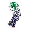
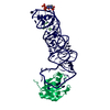
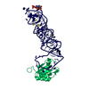
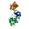
| Title | Structure of a c-di-GMP riboswitch from V. cholerae | ||||||
 Components Components |
| ||||||
 Keywords Keywords | RNA binding protein/RNA /  riboswitch / c-di-GMP / RNA / RNA binding protein-RNA complex riboswitch / c-di-GMP / RNA / RNA binding protein-RNA complex | ||||||
| Function / homology |  Function and homology information Function and homology informationU1 snRNP binding / U1 snRNP / U1 snRNA binding / U4/U6 x U5 tri-snRNP complex / mRNA Splicing - Major Pathway / spliceosomal complex / mRNA splicing, via spliceosome / DNA binding / RNA binding / nucleoplasm ...U1 snRNP binding / U1 snRNP / U1 snRNA binding / U4/U6 x U5 tri-snRNP complex / mRNA Splicing - Major Pathway / spliceosomal complex / mRNA splicing, via spliceosome / DNA binding / RNA binding / nucleoplasm / identical protein binding / nucleusSimilarity search - Function | ||||||
| Biological species |  Homo sapiens (human) Homo sapiens (human)  Vibrio cholerae (bacteria) Vibrio cholerae (bacteria) | ||||||
| Method | X-RAY DIFFRACTION / SYNCHROTRON / MAD / Resolution: 2.7 Å | ||||||
 Authors Authors | Smith, K.D. | ||||||
 Citation Citation | Journal: Nat.Struct.Mol.Biol. / Year: 2009 Title: Structural basis of ligand binding by a c-di-GMP riboswitch. Authors: Smith, K.D. / Lipchock, S.V. / Ames, T.D. / Wang, J. / Breaker, R.R. / Strobel, S.A. | ||||||
| History |
|
- Structure visualization
| Structure viewer | Molecule: MolmilJmol/JSmol |
|---|
- Downloads & links
-Download
| PDBx/mmCIF format | 3irw.cif.gz | 90.4 KB | Display | PDBx/mmCIF format |
|---|---|---|---|---|
| PDB format | pdb3irw.ent.gz | 64.8 KB | Display | PDB format |
| PDBx/mmJSON format | 3irw.json.gz | Tree view | PDBx/mmJSON format | |
| Others |  Other downloads Other downloads |
-Validation report
| Arichive directory | https://data.pdbj.org/pub/pdb/validation_reports/ir/3irwftp://data.pdbj.org/pub/pdb/validation_reports/ir/3irw | HTTPS FTP |
|---|
-Related structure data
| Similar structure data |
|---|


-Links
 PDBj
PDBj


- Assembly
| Deposited unit | 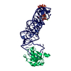
| ||||||||
|---|---|---|---|---|---|---|---|---|---|
| 1 |
| ||||||||
| Unit cell |
|
-Components
-Protein / RNA chain , 2 types, 2 molecules PR
| #1: Protein | Mass: 11340.315 Da / Num. of mol.: 1 / Fragment: RNA Binding Domain / Mutation: Y31H, Q36R Source method: isolated from a genetically manipulated source Source: (gene. exp.) Homo sapiens (human) / Gene: SNRPA / Plasmid: pET11 / Production host: Escherichia coli (E. coli) / Strain (production host): Bl21 / References: UniProt: P09012 |
|---|---|
| #2: RNA chain | Mass: 29942.773 Da / Num. of mol.: 1 / Source method: obtained synthetically Details: In vitro synthesis from a plasmid dna template of natural sequence from vibrio cholerae Source: (synth.) Vibrio cholerae (bacteria) |
-Non-polymers , 4 types, 104 molecules 


| #3: Chemical | ChemComp-C2E / Cyclic di-GMP Mass: 690.411 Da / Num. of mol.: 1 / Source method: obtained synthetically / Formula: C20H24N10O14P2 Mass: 690.411 Da / Num. of mol.: 1 / Source method: obtained synthetically / Formula: C20H24N10O14P2 | ||||
|---|---|---|---|---|---|
| #4: Chemical | ChemComp-IRI /  Mass: 294.400 Da / Num. of mol.: 9 / Source method: obtained synthetically / Formula: H18IrN6 Mass: 294.400 Da / Num. of mol.: 9 / Source method: obtained synthetically / Formula: H18IrN6#5: Chemical |  Mass: 24.305 Da / Num. of mol.: 2 / Source method: obtained synthetically / Formula: Mg Mass: 24.305 Da / Num. of mol.: 2 / Source method: obtained synthetically / Formula: Mg#6: Water | ChemComp-HOH / | WaterMass: 18.015 Da / Num. of mol.: 92 / Source method: isolated from a natural source / Formula: H2O |
-Experimental details
-Experiment
| Experiment | Method: X-RAY DIFFRACTION / Number of used crystals: 1 |
|---|
- Sample preparation
| Crystal | Density Matthews: 2.06 Å3/Da / Density % sol: 40.15 % |
|---|---|
| Crystal grow | Temperature: 298 K / pH: 6 Details: 22% PEG550mme, 50 mM MES, pH 6.0, 5 mM MgSO4, 300 mM NaCl, VAPOR DIFFUSION, HANGING DROP, temperature 298K |
-Data collection
| Diffraction | Mean temperature: 100 K | ||||||||||||
|---|---|---|---|---|---|---|---|---|---|---|---|---|---|
| Diffraction source | Source: SYNCHROTRON / Site: NSLS  / Beamline: X29A / Wavelength: 1.1050, 1.1054, 1.0762 / Beamline: X29A / Wavelength: 1.1050, 1.1054, 1.0762 | ||||||||||||
| Detector | Type: ADSC QUANTUM 315 / Detector: CCD / Date: Jan 26, 2009 Details: CRYOGENICALLY COOLED DOUBLE CRYSTAL MONOCHROMETER WITH HORIZONTAL FOCUSING SAGITTAL BEND SECOND MONO CRYSTAL WITH 4:1 MAGNIFICATION RATIO AND VERTICALLY FOCUSING MIRROR. | ||||||||||||
| Radiation | Monochromator: DOUBLE CRYSTAL MONOCHROMETER WITH HORIZONTAL FOCUSING SAGITTAL BEND SECOND MONO CRYSTAL WITH 4:1 MAGNIFICATION RATIO AND VERTICALLY FOCUSING MIRROR. Protocol: MAD / Monochromatic (M) / Laue (L): M / Scattering type: x-ray | ||||||||||||
| Radiation wavelength |
| ||||||||||||
| Reflection | Resolution: 2.7→50 Å / Num. obs: 8935 / % possible obs: 94.8 % / Observed criterion σ(I): 1.9 / Redundancy: 2.7 % / Rmerge(I) obs: 0.072 / Net I/σ(I): 12.5 | ||||||||||||
| Reflection shell | Resolution: 2.7→2.8 Å / Redundancy: 2 % / Rmerge(I) obs: 0.382 / Mean I/σ(I) obs: 1.9 / % possible all: 82.4 |
- Processing
| Software |
| ||||||||||||||||||||||||||||||||||||||||||||||||||||||||||||||||||||||||||||||||||||||||||||||||||||||||||||||||||||||||||||||||||||||||||||||||||||||||||||||||||||||||||
|---|---|---|---|---|---|---|---|---|---|---|---|---|---|---|---|---|---|---|---|---|---|---|---|---|---|---|---|---|---|---|---|---|---|---|---|---|---|---|---|---|---|---|---|---|---|---|---|---|---|---|---|---|---|---|---|---|---|---|---|---|---|---|---|---|---|---|---|---|---|---|---|---|---|---|---|---|---|---|---|---|---|---|---|---|---|---|---|---|---|---|---|---|---|---|---|---|---|---|---|---|---|---|---|---|---|---|---|---|---|---|---|---|---|---|---|---|---|---|---|---|---|---|---|---|---|---|---|---|---|---|---|---|---|---|---|---|---|---|---|---|---|---|---|---|---|---|---|---|---|---|---|---|---|---|---|---|---|---|---|---|---|---|---|---|---|---|---|---|---|---|---|
| Refinement | Method to determine structure: MAD / Resolution: 2.7→43.69 Å / Cor.coef. Fo:Fc: 0.952 / Cor.coef. Fo:Fc free: 0.923 / SU ML: 0.32 / TLS residual ADP flag: UNVERIFIED / Cross valid method: THROUGHOUT / ESU R Free: 0.411 Stereochemistry target values: MAXIMUM LIKELIHOOD WITH PHASES Details: HYDROGENS HAVE BEEN ADDED IN THE RIDING POSITIONS
| ||||||||||||||||||||||||||||||||||||||||||||||||||||||||||||||||||||||||||||||||||||||||||||||||||||||||||||||||||||||||||||||||||||||||||||||||||||||||||||||||||||||||||
| Solvent computation | Ion probe radii: 0.8 Å / Shrinkage radii: 0.8 Å / VDW probe radii: 1.2 Å / Solvent model: MASK | ||||||||||||||||||||||||||||||||||||||||||||||||||||||||||||||||||||||||||||||||||||||||||||||||||||||||||||||||||||||||||||||||||||||||||||||||||||||||||||||||||||||||||
| Displacement parameters | Biso mean: 50.651 Å2
| ||||||||||||||||||||||||||||||||||||||||||||||||||||||||||||||||||||||||||||||||||||||||||||||||||||||||||||||||||||||||||||||||||||||||||||||||||||||||||||||||||||||||||
| Refinement step | Cycle: LAST / Resolution: 2.7→43.69 Å
| ||||||||||||||||||||||||||||||||||||||||||||||||||||||||||||||||||||||||||||||||||||||||||||||||||||||||||||||||||||||||||||||||||||||||||||||||||||||||||||||||||||||||||
| Refine LS restraints |
| ||||||||||||||||||||||||||||||||||||||||||||||||||||||||||||||||||||||||||||||||||||||||||||||||||||||||||||||||||||||||||||||||||||||||||||||||||||||||||||||||||||||||||
| LS refinement shell | Resolution: 2.7→2.77 Å / Total num. of bins used: 20
| ||||||||||||||||||||||||||||||||||||||||||||||||||||||||||||||||||||||||||||||||||||||||||||||||||||||||||||||||||||||||||||||||||||||||||||||||||||||||||||||||||||||||||
| Refinement TLS params. | Method: refined / Refine-ID: X-RAY DIFFRACTION
| ||||||||||||||||||||||||||||||||||||||||||||||||||||||||||||||||||||||||||||||||||||||||||||||||||||||||||||||||||||||||||||||||||||||||||||||||||||||||||||||||||||||||||
| Refinement TLS group |
|
