 Movie
Movie Controller

[English] 日本語
 Yorodumi
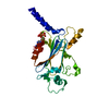
- PDB-3ilz: Structure of TR-alfa bound to selective thyromimetic GC-1 in P212... -
+ Open data

- Basic information
| Entry | Database: PDB / ID: 3ilz | ||||||
|---|---|---|---|---|---|---|---|
| Title | Structure of TR-alfa bound to selective thyromimetic GC-1 in P212121 space group | ||||||
 Components Components | Thyroid hormone receptor, alpha isoform 1 variant | ||||||
 Keywords Keywords | SIGNALING PROTEIN / nuclear receptor | ||||||
| Function / homology |  Function and homology information Function and homology informationregulation of myeloid cell apoptotic process / negative regulation of DNA-templated transcription initiation / female courtship behavior / negative regulation of RNA polymerase II transcription preinitiation complex assembly / regulation of lipid catabolic process / thyroid hormone mediated signaling pathway / positive regulation of thyroid hormone mediated signaling pathway / regulation of thyroid hormone mediated signaling pathway / positive regulation of female receptivity / regulation of heart contraction ...regulation of myeloid cell apoptotic process / negative regulation of DNA-templated transcription initiation / female courtship behavior / negative regulation of RNA polymerase II transcription preinitiation complex assembly / regulation of lipid catabolic process / thyroid hormone mediated signaling pathway / positive regulation of thyroid hormone mediated signaling pathway / regulation of thyroid hormone mediated signaling pathway / positive regulation of female receptivity / regulation of heart contraction / cartilage condensation / type I pneumocyte differentiation / thyroid hormone binding / thyroid gland development / general transcription initiation factor binding / retinoic acid receptor signaling pathway / TBP-class protein binding / response to cold / hormone-mediated signaling pathway / ossification / erythrocyte differentiation / SUMOylation of intracellular receptors / mRNA transcription by RNA polymerase II / chromatin DNA binding / Nuclear Receptor transcription pathway / RNA polymerase II transcription regulator complex / nuclear receptor activity / positive regulation of cold-induced thermogenesis / sequence-specific DNA binding / transcription by RNA polymerase II / learning or memory / cell differentiation / transcription cis-regulatory region binding / DNA-binding transcription factor activity, RNA polymerase II-specific / RNA polymerase II cis-regulatory region sequence-specific DNA binding / DNA-binding transcription factor activity / protein domain specific binding / negative regulation of DNA-templated transcription / chromatin / regulation of transcription by RNA polymerase II / negative regulation of transcription by RNA polymerase II / positive regulation of transcription by RNA polymerase II / zinc ion binding / nucleoplasm / nucleus / cytosolSimilarity search - Function | ||||||
| Biological species |  Homo sapiens (human) Homo sapiens (human) | ||||||
| Method | X-RAY DIFFRACTION / MOLECULAR REPLACEMENT / Resolution: 1.85 Å | ||||||
 Authors Authors | Aparicio, R. / Polikarpov, L. / Bleicher, L. | ||||||
 Citation Citation | Journal: Bmc Struct.Biol. / Year: 2008 Title: Structural basis of GC-1 selectivity for thyroid hormone receptor isoforms. Authors: Bleicher, L. / Aparicio, R. / Nunes, F.M. / Martinez, L. / Gomes Dias, S.M. / Figueira, A.C. / Santos, M.A. / Venturelli, W.H. / da Silva, R. / Donate, P.M. / Neves, F.A. / Simeoni, L.A. / ...Authors: Bleicher, L. / Aparicio, R. / Nunes, F.M. / Martinez, L. / Gomes Dias, S.M. / Figueira, A.C. / Santos, M.A. / Venturelli, W.H. / da Silva, R. / Donate, P.M. / Neves, F.A. / Simeoni, L.A. / Baxter, J.D. / Webb, P. / Skaf, M.S. / Polikarpov, I. | ||||||
| History |
|
- Structure visualization
| Structure viewer | Molecule: MolmilJmol/JSmol |
|---|
- Downloads & links
-Download
| PDBx/mmCIF format | 3ilz.cif.gz | 82.2 KB | Display | PDBx/mmCIF format |
|---|---|---|---|---|
| PDB format | pdb3ilz.ent.gz | 59.7 KB | Display | PDB format |
| PDBx/mmJSON format | 3ilz.json.gz | Tree view | PDBx/mmJSON format | |
| Others |  Other downloads Other downloads |
-Validation report
| Arichive directory | https://data.pdbj.org/pub/pdb/validation_reports/il/3ilzftp://data.pdbj.org/pub/pdb/validation_reports/il/3ilz | HTTPS FTP |
|---|
-Related structure data
| Related structure data |  3hzfC 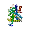 3imyC  1bsxS S: Starting model for refinement C: citing same article ( |
|---|---|
| Similar structure data |


-Links
 PDBj
PDBj
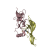
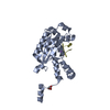
- Assembly
| Deposited unit | 
| ||||||||
|---|---|---|---|---|---|---|---|---|---|
| 1 |
| ||||||||
| Unit cell |
|
-Components
| #1: Protein | Mass: 30777.945 Da / Num. of mol.: 1 Source method: isolated from a genetically manipulated source Source: (gene. exp.) Homo sapiens (human) / Gene: NR1A1 / Plasmid: pET28a / Production host:  Escherichia coli (E. coli) / Strain (production host): B834 / References: UniProt: Q59FW3, UniProt: P10827*PLUS Escherichia coli (E. coli) / Strain (production host): B834 / References: UniProt: Q59FW3, UniProt: P10827*PLUS |
|---|---|
| #2: Chemical | ChemComp-B72 / {  Mass: 328.402 Da / Num. of mol.: 1 / Source method: obtained synthetically / Formula: C20H24O4 Mass: 328.402 Da / Num. of mol.: 1 / Source method: obtained synthetically / Formula: C20H24O4 |
| #3: Water | ChemComp-HOH / Water Mass: 18.015 Da / Num. of mol.: 477 / Source method: isolated from a natural source / Formula: H2O Mass: 18.015 Da / Num. of mol.: 477 / Source method: isolated from a natural source / Formula: H2O |
-Experimental details
-Experiment
| Experiment | Method: X-RAY DIFFRACTION / Number of used crystals: 1 |
|---|
- Sample preparation
| Crystal | Density Matthews: 4.02 Å3/Da / Density % sol: 69.43 % |
|---|---|
| Crystal grow | Temperature: 291 K / Method: vapor diffusion / pH: 7.2 Details: 1.0M NaCac 0.1M NaH3OAc, pH 7.2, VAPOR DIFFUSION, temperature 291K |
-Data collection
| Diffraction | Mean temperature: 100 K |
|---|---|
| Diffraction source | Source: ROTATING ANODE / Type: RIGAKU ULTRAX 18 / Wavelength: 1.5418 Å |
| Detector | Type: MAR scanner 345 mm plate / Detector: IMAGE PLATE / Date: Jan 1, 2004 |
| Radiation | Monochromator: OSMIC / Protocol: SINGLE WAVELENGTH / Monochromatic (M) / Laue (L): M / Scattering type: x-ray |
| Radiation wavelength | Wavelength: 1.5418 Å / Relative weight: 1 |
| Reflection | Resolution: 1.85→31.47 Å / Num. obs: 47773 / % possible obs: 98.7 % / Observed criterion σ(F): 1 / Observed criterion σ(I): 1 / Redundancy: 8.2 % / Rmerge(I) obs: 0.059 / Rsym value: 0.059 / Net I/σ(I): 7.8 |
| Reflection shell | Resolution: 1.85→1.898 Å / Redundancy: 7.8 % / Rmerge(I) obs: 0.383 / Mean I/σ(I) obs: 2 / Num. unique all: 2964 / Rsym value: 0.383 / % possible all: 98.9 |
- Processing
| Software |
| ||||||||||||||||||||||||||||||||||||||||||||||||||||||||||||||||||||||||||||||||||||||||||||||||||||
|---|---|---|---|---|---|---|---|---|---|---|---|---|---|---|---|---|---|---|---|---|---|---|---|---|---|---|---|---|---|---|---|---|---|---|---|---|---|---|---|---|---|---|---|---|---|---|---|---|---|---|---|---|---|---|---|---|---|---|---|---|---|---|---|---|---|---|---|---|---|---|---|---|---|---|---|---|---|---|---|---|---|---|---|---|---|---|---|---|---|---|---|---|---|---|---|---|---|---|---|---|---|
| Refinement | Method to determine structure: MOLECULAR REPLACEMENT Starting model: 1BSX Resolution: 1.85→31.47 Å / Cor.coef. Fo:Fc: 0.972 / Cor.coef. Fo:Fc free: 0.957 / Occupancy max: 1 / Occupancy min: 0 / SU B: 1.763 / SU ML: 0.054 / Cross valid method: THROUGHOUT / σ(F): 0 / ESU R: 0.083 / ESU R Free: 0.09 / Stereochemistry target values: MAXIMUM LIKELIHOOD / Details: HYDROGENS HAVE BEEN ADDED IN THE RIDING POSITIONS
| ||||||||||||||||||||||||||||||||||||||||||||||||||||||||||||||||||||||||||||||||||||||||||||||||||||
| Solvent computation | Ion probe radii: 0.8 Å / Shrinkage radii: 0.8 Å / VDW probe radii: 1.4 Å / Solvent model: BABINET MODEL WITH MASK | ||||||||||||||||||||||||||||||||||||||||||||||||||||||||||||||||||||||||||||||||||||||||||||||||||||
| Displacement parameters | Biso max: 95.41 Å2 / Biso mean: 29.418 Å2 / Biso min: 2 Å2
| ||||||||||||||||||||||||||||||||||||||||||||||||||||||||||||||||||||||||||||||||||||||||||||||||||||
| Refinement step | Cycle: LAST / Resolution: 1.85→31.47 Å
| ||||||||||||||||||||||||||||||||||||||||||||||||||||||||||||||||||||||||||||||||||||||||||||||||||||
| Refine LS restraints |
| ||||||||||||||||||||||||||||||||||||||||||||||||||||||||||||||||||||||||||||||||||||||||||||||||||||
| LS refinement shell | Resolution: 1.85→1.898 Å / Total num. of bins used: 20
| ||||||||||||||||||||||||||||||||||||||||||||||||||||||||||||||||||||||||||||||||||||||||||||||||||||
| Refinement TLS params. | Method: refined / Refine-ID: X-RAY DIFFRACTION
| ||||||||||||||||||||||||||||||||||||||||||||||||||||||||||||||||||||||||||||||||||||||||||||||||||||
| Refinement TLS group |
|
