 Movie
Movie Controller

[English] 日本語
 Yorodumi
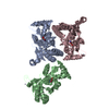
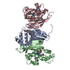
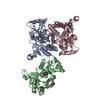
- PDB-3ilt: Crystal structure of the AMPA subunit GluR2 bound to the alloster... -
+ Open data

- Basic information
| Entry | Database: PDB / ID: 3ilt | ||||||
|---|---|---|---|---|---|---|---|
| Title | Crystal structure of the AMPA subunit GluR2 bound to the allosteric modulator, trichlormethiazide | ||||||
 Components Components | Glutamate receptor 2 GRIA2 GRIA2 | ||||||
 Keywords Keywords | SIGNALING PROTEIN / glutamate receptor / glur2 / AMPA receptor / neurotransmitter receptor / S1S2 / allosteric modulator | ||||||
| Function / homology |  Function and homology information Function and homology informationspine synapse / dendritic spine neck / dendritic spine head / Activation of AMPA receptors / response to lithium ion / perisynaptic space / cellular response to glycine / AMPA glutamate receptor activity / Trafficking of GluR2-containing AMPA receptors / immunoglobulin binding ...spine synapse / dendritic spine neck / dendritic spine head / Activation of AMPA receptors / response to lithium ion / perisynaptic space / cellular response to glycine / AMPA glutamate receptor activity / Trafficking of GluR2-containing AMPA receptors / immunoglobulin binding / AMPA glutamate receptor complex / kainate selective glutamate receptor activity / ionotropic glutamate receptor complex / extracellularly glutamate-gated ion channel activity / asymmetric synapse / regulation of receptor recycling / Unblocking of NMDA receptors, glutamate binding and activation / glutamate receptor binding / positive regulation of synaptic transmission / glutamate-gated receptor activity / presynaptic active zone membrane / response to fungicide / regulation of synaptic transmission, glutamatergic / cellular response to brain-derived neurotrophic factor stimulus / somatodendritic compartment / dendrite membrane / ligand-gated monoatomic ion channel activity involved in regulation of presynaptic membrane potential / ionotropic glutamate receptor binding / ionotropic glutamate receptor signaling pathway / dendrite cytoplasm / cytoskeletal protein binding / SNARE binding / dendritic shaft / transmitter-gated monoatomic ion channel activity involved in regulation of postsynaptic membrane potential / synaptic membrane / synaptic transmission, glutamatergic / PDZ domain binding / postsynaptic density membrane / protein tetramerization / modulation of chemical synaptic transmission / Schaffer collateral - CA1 synapse / establishment of protein localization / terminal bouton / receptor internalization / synaptic vesicle membrane / cerebral cortex development / synaptic vesicle / presynapse / signaling receptor activity / presynaptic membrane / amyloid-beta binding / growth cone / perikaryon / chemical synaptic transmission / postsynaptic membrane / scaffold protein binding / dendritic spine / postsynaptic density / neuron projection / axon / dendrite / neuronal cell body / glutamatergic synapse / synapse / protein-containing complex binding / endoplasmic reticulum membrane / protein kinase binding / cell surface / endoplasmic reticulum / protein-containing complex / membrane / identical protein binding / plasma membraneSimilarity search - Function | ||||||
| Biological species |  Rattus norvegicus (Norway rat) Rattus norvegicus (Norway rat) | ||||||
| Method | X-RAY DIFFRACTION / SYNCHROTRON / MOLECULAR REPLACEMENT / molecular replacement / Resolution: 2.107 Å | ||||||
 Authors Authors | Ahmed, A.H. / Ptak, C.P. / Oswald, R.E. | ||||||
 Citation Citation | Journal: Biochemistry / Year: 2009 Title: Probing the allosteric modulator binding site of GluR2 with thiazide derivatives Authors: Ptak, C.P. / Ahmed, A.H. / Oswald, R.E. | ||||||
| History |
|
- Structure visualization
| Structure viewer | Molecule: MolmilJmol/JSmol |
|---|
- Downloads & links
-Download
| PDBx/mmCIF format | 3ilt.cif.gz | 163.4 KB | Display | PDBx/mmCIF format |
|---|---|---|---|---|
| PDB format | pdb3ilt.ent.gz | 128.2 KB | Display | PDB format |
| PDBx/mmJSON format | 3ilt.json.gz | Tree view | PDBx/mmJSON format | |
| Others |  Other downloads Other downloads |
-Validation report
| Arichive directory | https://data.pdbj.org/pub/pdb/validation_reports/il/3iltftp://data.pdbj.org/pub/pdb/validation_reports/il/3ilt | HTTPS FTP |
|---|
-Related structure data
| Related structure data |  3ijoC  3ijxC 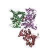 3ik6C  3il1C  3iluC  3dp6S C: citing same article ( S: Starting model for refinement |
|---|---|
| Similar structure data |


-Links
 PDBj
PDBj


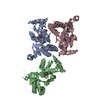
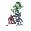
- Assembly
| Deposited unit | 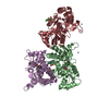
| ||||||||
|---|---|---|---|---|---|---|---|---|---|
| 1 |
| ||||||||
| Unit cell |
|
-Components
| #1: Protein | GRIA2 / GluR-2 / GluR-B / GluR-K2 / Glutamate receptor ionotropic / AMPA 2 / AMPA-selective glutamate receptor 2 Mass: 28808.297 Da / Num. of mol.: 3 / Fragment: S1S2 binding domain / Mutation: N242S Source method: isolated from a genetically manipulated source Source: (gene. exp.) Rattus norvegicus (Norway rat) / Gene: Glur2, Gria2, Gria2; GluR2 / Plasmid: pET-22b(+) / Production host:  Escherichia coli (E. coli) / Strain (production host): Origami B (DE3) / References: UniProt: P19491 Escherichia coli (E. coli) / Strain (production host): Origami B (DE3) / References: UniProt: P19491#2: Chemical | Glutamic acid  Type: L-peptide linking / Mass: 147.129 Da / Num. of mol.: 3 / Source method: obtained synthetically / Formula: C5H9NO4 Type: L-peptide linking / Mass: 147.129 Da / Num. of mol.: 3 / Source method: obtained synthetically / Formula: C5H9NO4#3: Chemical | ChemComp-ZN /   Mass: 65.409 Da / Num. of mol.: 5 / Source method: obtained synthetically / Formula: Zn Mass: 65.409 Da / Num. of mol.: 5 / Source method: obtained synthetically / Formula: Zn#4: Chemical | 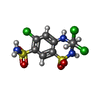  Mass: 380.656 Da / Num. of mol.: 3 / Source method: obtained synthetically / Formula: C8H8Cl3N3O4S2 Mass: 380.656 Da / Num. of mol.: 3 / Source method: obtained synthetically / Formula: C8H8Cl3N3O4S2#5: Water | ChemComp-HOH / | Water Mass: 18.015 Da / Num. of mol.: 153 / Source method: isolated from a natural source / Formula: H2O Mass: 18.015 Da / Num. of mol.: 153 / Source method: isolated from a natural source / Formula: H2O |
|---|
-Experimental details
-Experiment
| Experiment | Method: X-RAY DIFFRACTION / Number of used crystals: 1 |
|---|
- Sample preparation
| Crystal | Density Matthews: 2.23 Å3/Da / Density % sol: 44.72 % |
|---|---|
| Crystal grow | Temperature: 277 K / Method: vapor diffusion, hanging drop / pH: 6.5 Details: 16-18 PEG8K, 0.1 M Na Cacodylate, 0.1-0.15 zinc acetate, pH 6.5, vapor diffusion, hanging drop, temperature 277K |
-Data collection
| Diffraction | Mean temperature: 100 K |
|---|---|
| Diffraction source | Source: SYNCHROTRON / Site: CHESS  / Beamline: A1 / Wavelength: 0.977 Å / Beamline: A1 / Wavelength: 0.977 Å |
| Detector | Type: ADSC QUANTUM 210 / Detector: CCD / Date: Dec 23, 2008 |
| Radiation | Monochromator: RH COATED SI / Protocol: SINGLE WAVELENGTH / Monochromatic (M) / Laue (L): M / Scattering type: x-ray |
| Radiation wavelength | Wavelength: 0.977 Å / Relative weight: 1 |
| Reflection | Resolution: 2.2→50 Å / Num. obs: 43133 / % possible obs: 99.9 % / Observed criterion σ(F): 0 / Observed criterion σ(I): -3 / Redundancy: 5.1 % / Rmerge(I) obs: 0.11 / Rsym value: 0.11 / Χ2: 1.432 / Net I/σ(I): 6.3 |
| Reflection shell | Resolution: 2.2→2.24 Å / Redundancy: 5.1 % / Rmerge(I) obs: 0.618 / Mean I/σ(I) obs: 2.154 / Num. unique all: 2086 / Rsym value: 0.618 / Χ2: 0.88 / % possible all: 100 |
-Phasing
| Phasing | Method: molecular replacement |
|---|
- Processing
| Software |
| |||||||||||||||||||||||||||||||||||||||||||||||||||||||||||||||||||||||||||||||||||||||||||||||||||||||||
|---|---|---|---|---|---|---|---|---|---|---|---|---|---|---|---|---|---|---|---|---|---|---|---|---|---|---|---|---|---|---|---|---|---|---|---|---|---|---|---|---|---|---|---|---|---|---|---|---|---|---|---|---|---|---|---|---|---|---|---|---|---|---|---|---|---|---|---|---|---|---|---|---|---|---|---|---|---|---|---|---|---|---|---|---|---|---|---|---|---|---|---|---|---|---|---|---|---|---|---|---|---|---|---|---|---|---|
| Refinement | Method to determine structure: MOLECULAR REPLACEMENT Starting model: PDB ENTRY 3dp6 Resolution: 2.107→21.234 Å / Occupancy max: 1 / Occupancy min: 1 / FOM work R set: 0.8 / SU ML: 0.35 / Isotropic thermal model: Isotropic / Cross valid method: THROUGHOUT / σ(F): 0.83 / σ(I): 0 / Stereochemistry target values: ML
| |||||||||||||||||||||||||||||||||||||||||||||||||||||||||||||||||||||||||||||||||||||||||||||||||||||||||
| Solvent computation | Shrinkage radii: 0.9 Å / VDW probe radii: 1.11 Å / Solvent model: FLAT BULK SOLVENT MODEL / Bsol: 49.762 Å2 / ksol: 0.437 e/Å3 | |||||||||||||||||||||||||||||||||||||||||||||||||||||||||||||||||||||||||||||||||||||||||||||||||||||||||
| Displacement parameters | Biso max: 89.76 Å2 / Biso mean: 43.215 Å2 / Biso min: 6.18 Å2
| |||||||||||||||||||||||||||||||||||||||||||||||||||||||||||||||||||||||||||||||||||||||||||||||||||||||||
| Refinement step | Cycle: LAST / Resolution: 2.107→21.234 Å
| |||||||||||||||||||||||||||||||||||||||||||||||||||||||||||||||||||||||||||||||||||||||||||||||||||||||||
| Refine LS restraints |
| |||||||||||||||||||||||||||||||||||||||||||||||||||||||||||||||||||||||||||||||||||||||||||||||||||||||||
| LS refinement shell | Refine-ID: X-RAY DIFFRACTION / Total num. of bins used: 14
|
