 Movie
Movie Controller

[English] 日本語
 Yorodumi
- PDB-3ik2: Crystal Structure of a Glycoside Hydrolase Family 44 Endoglucanas... -
+ Open data

- Basic information
| Entry | Database: PDB / ID: 3ik2 | ||||||
|---|---|---|---|---|---|---|---|
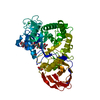
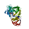
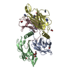
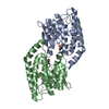
| Title | Crystal Structure of a Glycoside Hydrolase Family 44 Endoglucanase produced by Clostridium acetobutylium ATCC 824 | ||||||
 Components Components | Endoglucanase A | ||||||
 Keywords Keywords |  HYDROLASE / TIM-like barrel HYDROLASE / TIM-like barrel | ||||||
| Function / homology |  Function and homology information Function and homology informationpolysaccharide catabolic process / hydrolase activity, hydrolyzing O-glycosyl compounds / metal ion bindingSimilarity search - Function | ||||||
| Biological species |  Clostridium acetobutylicum (bacteria) Clostridium acetobutylicum (bacteria) | ||||||
| Method | X-RAY DIFFRACTION / MOLECULAR REPLACEMENT / molecular replacement / Resolution: 2.2 Å | ||||||
 Authors Authors | Warner, C.D. / Hoy, J.A. / Ford, C.F. / Honzatko, R.B. / Reilly, P.J. | ||||||
 Citation Citation | Journal: Appl.Environ.Microbiol. / Year: 2010 Title: Tertiary structure and characterization of a glycoside hydrolase family 44 endoglucanase from Clostridium acetobutylicum. Authors: Warner, C.D. / Hoy, J.A. / Shilling, T.C. / Linnen, M.J. / Ginder, N.D. / Ford, C.F. / Honzatko, R.B. / Reilly, P.J. | ||||||
| History |
|
- Structure visualization
| Structure viewer | Molecule: MolmilJmol/JSmol |
|---|
- Downloads & links
-Download
| PDBx/mmCIF format | 3ik2.cif.gz | 132.5 KB | Display | PDBx/mmCIF format |
|---|---|---|---|---|
| PDB format | pdb3ik2.ent.gz | 99.1 KB | Display | PDB format |
| PDBx/mmJSON format | 3ik2.json.gz | Tree view | PDBx/mmJSON format | |
| Others |  Other downloads Other downloads |
-Validation report
| Arichive directory | https://data.pdbj.org/pub/pdb/validation_reports/ik/3ik2ftp://data.pdbj.org/pub/pdb/validation_reports/ik/3ik2 | HTTPS FTP |
|---|
-Related structure data
| Related structure data |  2e4tS S: Starting model for refinement |
|---|---|
| Similar structure data |


-Links
 PDBj
PDBj- Assembly
| Deposited unit | 
| ||||||||
|---|---|---|---|---|---|---|---|---|---|
| 1 |
| ||||||||
| Unit cell |
|
-Components
-Protein , 1 types, 1 molecules A
| #1: Protein | Mass: 57653.953 Da / Num. of mol.: 1 Fragment: UNP RESIDUES 34-541(Fragment excluding signal peptide & dockerin domain) Source method: isolated from a genetically manipulated source Details: Gene synthesized by Megabase (Lincoln, NE) / Source: (gene. exp.) Clostridium acetobutylicum (bacteria) / Strain: ATCC 824 / Gene: CAC0915, CA_C0915 / Plasmid: pET-22b / Production host: Escherichia coli (E. coli) / Strain (production host): BL21(DE3) / References: UniProt: Q977Y3 |
|---|
-Non-polymers , 6 types, 653 molecules 


| #2: Chemical | ChemComp-CL / Chloride Mass: 35.453 Da / Num. of mol.: 1 / Source method: obtained synthetically / Formula: Cl Mass: 35.453 Da / Num. of mol.: 1 / Source method: obtained synthetically / Formula: Cl | ||||||
|---|---|---|---|---|---|---|---|
| #3: Chemical | ChemComp-CA /  Mass: 40.078 Da / Num. of mol.: 1 / Source method: obtained synthetically / Formula: Ca Mass: 40.078 Da / Num. of mol.: 1 / Source method: obtained synthetically / Formula: Ca | ||||||
| #4: Chemical | ChemComp-GOL / Glycerol Mass: 92.094 Da / Num. of mol.: 9 / Source method: obtained synthetically / Formula: C3H8O3 Mass: 92.094 Da / Num. of mol.: 9 / Source method: obtained synthetically / Formula: C3H8O3#5: Chemical | ChemComp-SO4 / | Sulfate Mass: 96.063 Da / Num. of mol.: 1 / Source method: obtained synthetically / Formula: SO4 Mass: 96.063 Da / Num. of mol.: 1 / Source method: obtained synthetically / Formula: SO4#6: Chemical | ChemComp-ACT / Acetate Mass: 59.044 Da / Num. of mol.: 4 / Source method: obtained synthetically / Formula: C2H3O2 Mass: 59.044 Da / Num. of mol.: 4 / Source method: obtained synthetically / Formula: C2H3O2#7: Water | ChemComp-HOH / | WaterMass: 18.015 Da / Num. of mol.: 637 / Source method: isolated from a natural source / Formula: H2O |
-Experimental details
-Experiment
| Experiment | Method: X-RAY DIFFRACTION / Number of used crystals: 1 |
|---|
- Sample preparation
| Crystal | Density Matthews: 2.14 Å3/Da / Density % sol: 42.51 % |
|---|---|
| Crystal grow | Temperature: 296 K / Method: vapor diffusion, hanging drop / pH: 5.4 Details: 0.2 M (NH4)2SO4, 0.1 M NaOAc, 10% (w/v) PEG 3,350, pH 5.4, vapor diffusion, hanging drop, temperature 296K |
-Data collection
| Diffraction | Mean temperature: 100 K |
|---|---|
| Diffraction source | Source: ROTATING ANODE / Type: RIGAKU / Wavelength: 1.54 Å |
| Detector | Type: RIGAKU RAXIS IV++ / Detector: IMAGE PLATE / Date: Nov 28, 2007 |
| Radiation | Monochromator: Tuned to copper / Protocol: SINGLE WAVELENGTH / Monochromatic (M) / Laue (L): M / Scattering type: x-ray |
| Radiation wavelength | Wavelength: 1.54 Å / Relative weight: 1 |
| Reflection | Resolution: 2.2→66.67 Å / Num. all: 25388 / Num. obs: 25388 / % possible obs: 99.5 % / Observed criterion σ(F): 5 / Observed criterion σ(I): 2 / Redundancy: 4.87 % / Rmerge(I) obs: 0.086 / Χ2: 0.96 / Net I/σ(I): 11.4 / Scaling rejects: 935 |
| Reflection shell | Resolution: 2.2→2.28 Å / Redundancy: 4.19 % / Rmerge(I) obs: 0.209 / Mean I/σ(I) obs: 5 / Num. measured all: 9990 / Num. unique all: 2363 / Χ2: 1.34 / % possible all: 94.3 |
-Phasing
| Phasing | Method: molecular replacement |
|---|
- Processing
| Software |
| |||||||||||||||||||||||||||||||||||||||||||||||||||||||||||||||||||||||||||||||||||||||||||||||
|---|---|---|---|---|---|---|---|---|---|---|---|---|---|---|---|---|---|---|---|---|---|---|---|---|---|---|---|---|---|---|---|---|---|---|---|---|---|---|---|---|---|---|---|---|---|---|---|---|---|---|---|---|---|---|---|---|---|---|---|---|---|---|---|---|---|---|---|---|---|---|---|---|---|---|---|---|---|---|---|---|---|---|---|---|---|---|---|---|---|---|---|---|---|---|---|---|
| Refinement | Method to determine structure: MOLECULAR REPLACEMENT Starting model: 2E4T.pdb Resolution: 2.2→66.67 Å / Cor.coef. Fo:Fc: 0.955 / Cor.coef. Fo:Fc free: 0.883 / Occupancy max: 1 / Occupancy min: 1 / SU B: 5.814 / SU ML: 0.151 / Cross valid method: THROUGHOUT / σ(F): 5 / σ(I): 2 / ESU R: 0.326 / ESU R Free: 0.236 / Stereochemistry target values: MAXIMUM LIKELIHOOD / Details: HYDROGENS HAVE BEEN ADDED IN THE RIDING POSITIONS
| |||||||||||||||||||||||||||||||||||||||||||||||||||||||||||||||||||||||||||||||||||||||||||||||
| Solvent computation | Ion probe radii: 0.8 Å / Shrinkage radii: 0.8 Å / VDW probe radii: 1.2 Å / Solvent model: MASK | |||||||||||||||||||||||||||||||||||||||||||||||||||||||||||||||||||||||||||||||||||||||||||||||
| Displacement parameters | Biso max: 243.86 Å2 / Biso mean: 13.764 Å2 / Biso min: 2 Å2
| |||||||||||||||||||||||||||||||||||||||||||||||||||||||||||||||||||||||||||||||||||||||||||||||
| Refinement step | Cycle: LAST / Resolution: 2.2→66.67 Å
| |||||||||||||||||||||||||||||||||||||||||||||||||||||||||||||||||||||||||||||||||||||||||||||||
| Refine LS restraints |
| |||||||||||||||||||||||||||||||||||||||||||||||||||||||||||||||||||||||||||||||||||||||||||||||
| LS refinement shell | Resolution: 2.2→2.257 Å / Total num. of bins used: 20
|
