 Movie
Movie Controller

[English] 日本語
 Yorodumi
- PDB-3igs: Structure of the Salmonella enterica N-acetylmannosamine-6-phosph... -
+ Open data

- Basic information
| Entry | Database: PDB / ID: 3igs | ||||||
|---|---|---|---|---|---|---|---|
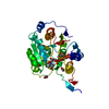
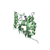
| Title | Structure of the Salmonella enterica N-acetylmannosamine-6-phosphate 2-epimerase | ||||||
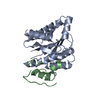
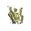
 Components Components | N-acetylmannosamine-6-phosphate 2-epimerase 2 | ||||||
 Keywords Keywords |  ISOMERASE / salmonella / energy metabolism / sugars / CSGID / Carbohydrate metabolism / Structural Genomics / Center for Structural Genomics of Infectious Diseases ISOMERASE / salmonella / energy metabolism / sugars / CSGID / Carbohydrate metabolism / Structural Genomics / Center for Structural Genomics of Infectious Diseases | ||||||
| Function / homology |  Function and homology information Function and homology informationN-acetylmannosamine catabolic process / N-acylglucosamine-6-phosphate 2-epimerase / N-acylmannosamine-6-phosphate 2-epimerase activity / N-acylglucosamine-6-phosphate 2-epimerase activity / N-acetylneuraminate catabolic process / carbohydrate metabolic process / cytosolSimilarity search - Function | ||||||
| Biological species |  Salmonella enterica subsp. enterica serovar Typhimurium (bacteria) Salmonella enterica subsp. enterica serovar Typhimurium (bacteria) | ||||||
| Method | X-RAY DIFFRACTION / SYNCHROTRON / SAD / Resolution: 1.5 Å | ||||||
 Authors Authors | Anderson, S.M. / Wawrzak, Z. / Gordon, E. / Skarina, T. / Papazisi, L. / Anderson, W.F. / Savchenko, A. / Center for Structural Genomics of Infectious Diseases (CSGID) | ||||||
| History |
|
- Structure visualization
| Structure viewer | Molecule: MolmilJmol/JSmol |
|---|
- Downloads & links
-Download
| PDBx/mmCIF format | 3igs.cif.gz | 119.1 KB | Display | PDBx/mmCIF format |
|---|---|---|---|---|
| PDB format | pdb3igs.ent.gz | 95.7 KB | Display | PDB format |
| PDBx/mmJSON format | 3igs.json.gz | Tree view | PDBx/mmJSON format | |
| Others |  Other downloads Other downloads |
-Validation report
| Arichive directory | https://data.pdbj.org/pub/pdb/validation_reports/ig/3igsftp://data.pdbj.org/pub/pdb/validation_reports/ig/3igs | HTTPS FTP |
|---|
-Related structure data
| Similar structure data | |
|---|---|
| Other databases |
-Links
 PDBj
PDBj
- Assembly
| Deposited unit | 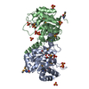
| ||||||||
|---|---|---|---|---|---|---|---|---|---|
| 1 | 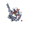
| ||||||||
| 2 | 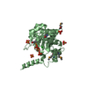
| ||||||||
| Unit cell |
|
-Components
| #1: Protein | Mass: 24591.344 Da / Num. of mol.: 2 / Mutation: A129T cloning error Source method: isolated from a genetically manipulated source Source: (gene. exp.) Salmonella enterica subsp. enterica serovar Typhimurium (bacteria)Strain: LT2 / Gene: nanE2, STM3337 / Production host: Escherichia coli (E. coli) / Strain (production host): BL21(DE3)References: UniProt: Q8ZLQ7, N-acylglucosamine-6-phosphate 2-epimerase#2: Sugar | ChemComp-16G / | 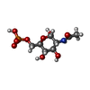  Type: D-saccharide, alpha linking / Mass: 301.188 Da / Num. of mol.: 1 Type: D-saccharide, alpha linking / Mass: 301.188 Da / Num. of mol.: 1Source method: isolated from a genetically manipulated source Formula: C8H16NO9P #3: Chemical | ChemComp-SO4 / Sulfate  Mass: 96.063 Da / Num. of mol.: 14 / Source method: obtained synthetically / Formula: SO4 Mass: 96.063 Da / Num. of mol.: 14 / Source method: obtained synthetically / Formula: SO4#4: Chemical | Chloride  Mass: 35.453 Da / Num. of mol.: 2 / Source method: obtained synthetically / Formula: Cl Mass: 35.453 Da / Num. of mol.: 2 / Source method: obtained synthetically / Formula: Cl#5: Water | ChemComp-HOH / | Water Mass: 18.015 Da / Num. of mol.: 878 / Source method: isolated from a natural source / Formula: H2O Mass: 18.015 Da / Num. of mol.: 878 / Source method: isolated from a natural source / Formula: H2O |
|---|
-Experimental details
-Experiment
| Experiment | Method: X-RAY DIFFRACTION / Number of used crystals: 1 |
|---|
- Sample preparation
| Crystal | Density Matthews: 2.47 Å3/Da / Density % sol: 50.11 % |
|---|---|
| Crystal grow | Temperature: 295 K / Method: vapor diffusion, hanging drop / pH: 7 Details: 2M ammonium sulphate, 5% isopropanol, pH 7.0, VAPOR DIFFUSION, HANGING DROP, temperature 295K |
-Data collection
| Diffraction | Mean temperature: 110 K |
|---|---|
| Diffraction source | Source: SYNCHROTRON / Site: APS  / Beamline: 21-ID-F / Wavelength: 0.97872 Å / Beamline: 21-ID-F / Wavelength: 0.97872 Å |
| Detector | Type: MARMOSAIC 300 mm CCD / Detector: CCD / Date: Jun 11, 2009 / Details: beryllium lense |
| Radiation | Monochromator: C(111) diamond laue monochromator / Protocol: SINGLE WAVELENGTH / Monochromatic (M) / Laue (L): M / Scattering type: x-ray |
| Radiation wavelength | Wavelength: 0.97872 Å / Relative weight: 1 |
| Reflection | Resolution: 1.5→50 Å / Num. all: 77012 / Num. obs: 74625 / % possible obs: 96.9 % / Observed criterion σ(F): 1.8 / Observed criterion σ(I): 3.3 / Redundancy: 7.6 % / Rmerge(I) obs: 0.095 / Χ2: 1.008 / Net I/σ(I): 26.7 |
| Reflection shell | Resolution: 1.5→1.55 Å / Redundancy: 4.7 % / Rmerge(I) obs: 0.393 / Mean I/σ(I) obs: 3.33 / Num. unique all: 5711 / Χ2: 0.979 / % possible all: 74.8 |
- Processing
| Software |
| |||||||||||||||||||||||||||||||||||||||||||||||||||||||||||||||||||||||||||
|---|---|---|---|---|---|---|---|---|---|---|---|---|---|---|---|---|---|---|---|---|---|---|---|---|---|---|---|---|---|---|---|---|---|---|---|---|---|---|---|---|---|---|---|---|---|---|---|---|---|---|---|---|---|---|---|---|---|---|---|---|---|---|---|---|---|---|---|---|---|---|---|---|---|---|---|---|
| Refinement | Method to determine structure: SAD / Resolution: 1.5→37.7 Å / Cor.coef. Fo:Fc: 0.969 / Cor.coef. Fo:Fc free: 0.957 / WRfactor Rfree: 0.187 / WRfactor Rwork: 0.156 / Occupancy max: 1 / Occupancy min: 0 / FOM work R set: 0.914 / SU B: 2.164 / SU ML: 0.038 / SU R Cruickshank DPI: 0.067 / SU Rfree: 0.069 / Cross valid method: THROUGHOUT / σ(F): 0 / ESU R: 0.067 / ESU R Free: 0.069 / Stereochemistry target values: MAXIMUM LIKELIHOOD Details: HYDROGENS HAVE BEEN ADDED IN THE RIDING POSITIONS U VALUES: RESIDUAL ONLY
| |||||||||||||||||||||||||||||||||||||||||||||||||||||||||||||||||||||||||||
| Solvent computation | Ion probe radii: 0.8 Å / Shrinkage radii: 0.8 Å / VDW probe radii: 1.4 Å / Solvent model: MASK | |||||||||||||||||||||||||||||||||||||||||||||||||||||||||||||||||||||||||||
| Displacement parameters | Biso max: 89.52 Å2 / Biso mean: 10.801 Å2 / Biso min: 2 Å2
| |||||||||||||||||||||||||||||||||||||||||||||||||||||||||||||||||||||||||||
| Refinement step | Cycle: LAST / Resolution: 1.5→37.7 Å
| |||||||||||||||||||||||||||||||||||||||||||||||||||||||||||||||||||||||||||
| Refine LS restraints |
| |||||||||||||||||||||||||||||||||||||||||||||||||||||||||||||||||||||||||||
| LS refinement shell | Resolution: 1.5→1.54 Å / Total num. of bins used: 20
| |||||||||||||||||||||||||||||||||||||||||||||||||||||||||||||||||||||||||||
| Refinement TLS params. | Method: refined / Refine-ID: X-RAY DIFFRACTION
| |||||||||||||||||||||||||||||||||||||||||||||||||||||||||||||||||||||||||||
| Refinement TLS group |
|
