 Movie
Movie Controller

[English] 日本語
 Yorodumi
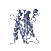
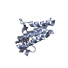
- PDB-3ifk: Crystal Structure Of Calcium-Saturated Calmodulin N-terminal Doma... -
+ Open data

- Basic information
| Entry | Database: PDB / ID: 3ifk | ||||||
|---|---|---|---|---|---|---|---|
| Title | Crystal Structure Of Calcium-Saturated Calmodulin N-terminal Domain Fragment, Residues 1-90 | ||||||
 Components Components | CALMODULIN | ||||||
 Keywords Keywords | METAL BINDING PROTEIN / CALMODULIN / EF HAND MOTIF / N-TERMINAL DOMAIN / N-DOMAIN / RESIDUES 1-90 / METHYLATION / PHOSPHORYLATION / Isopeptide bond / Phosphoprotein | ||||||
| Function / homology |  Function and homology informationregulation of store-operated calcium channel activity / regulation of high voltage-gated calcium channel activity / : / regulation of response to tumor cell / positive regulation of autophagic cell death / DAPK1-calmodulin complex / : / establishment of protein localization to mitochondrial membrane / type 3 metabotropic glutamate receptor binding / establishment of protein localization to membrane ...regulation of store-operated calcium channel activity / regulation of high voltage-gated calcium channel activity / : / regulation of response to tumor cell / positive regulation of autophagic cell death / DAPK1-calmodulin complex / : / establishment of protein localization to mitochondrial membrane / type 3 metabotropic glutamate receptor binding / establishment of protein localization to membrane / regulation of synaptic vesicle endocytosis / negative regulation of high voltage-gated calcium channel activity / regulation of synaptic vesicle exocytosis / organelle localization by membrane tethering / negative regulation of calcium ion export across plasma membrane / mitochondrion-endoplasmic reticulum membrane tethering / regulation of cardiac muscle cell action potential / autophagosome membrane docking / positive regulation of ryanodine-sensitive calcium-release channel activity / nitric-oxide synthase binding / protein phosphatase activator activity / positive regulation of cyclic-nucleotide phosphodiesterase activity / positive regulation of phosphoprotein phosphatase activity / adenylate cyclase binding / catalytic complex / detection of calcium ion / calcium channel regulator activity / negative regulation of ryanodine-sensitive calcium-release channel activity / regulation of cardiac muscle contraction / calcium channel inhibitor activity / cellular response to interferon-beta / positive regulation of DNA binding / phosphatidylinositol 3-kinase binding / enzyme regulator activity / regulation of release of sequestered calcium ion into cytosol by sarcoplasmic reticulum / regulation of calcium-mediated signaling / positive regulation of protein dephosphorylation / potassium ion transmembrane transport / voltage-gated potassium channel complex / regulation of ryanodine-sensitive calcium-release channel activity / titin binding / sperm midpiece / calcium channel complex / activation of adenylate cyclase activity / response to amphetamine / adenylate cyclase activator activity / nitric-oxide synthase regulator activity / regulation of heart rate / sarcomere / protein serine/threonine kinase activator activity / regulation of cytokinesis / calcium-mediated signaling / positive regulation of nitric-oxide synthase activity / positive regulation of receptor signaling pathway via JAK-STAT / spindle microtubule / cellular response to type II interferon / spindle pole / response to calcium ion / calcium-dependent protein binding / G2/M transition of mitotic cell cycle / disordered domain specific binding / myelin sheath / growth cone / vesicle / transmembrane transporter binding / protein autophosphorylation / neuron projection / positive regulation of apoptotic process / protein domain specific binding / centrosome / calcium ion binding / protein kinase binding / protein-containing complex / mitochondrion / nucleoplasm / nucleus / plasma membrane / cytosol / cytoplasm Function and homology informationregulation of store-operated calcium channel activity / regulation of high voltage-gated calcium channel activity / : / regulation of response to tumor cell / positive regulation of autophagic cell death / DAPK1-calmodulin complex / : / establishment of protein localization to mitochondrial membrane / type 3 metabotropic glutamate receptor binding / establishment of protein localization to membrane ...regulation of store-operated calcium channel activity / regulation of high voltage-gated calcium channel activity / : / regulation of response to tumor cell / positive regulation of autophagic cell death / DAPK1-calmodulin complex / : / establishment of protein localization to mitochondrial membrane / type 3 metabotropic glutamate receptor binding / establishment of protein localization to membrane / regulation of synaptic vesicle endocytosis / negative regulation of high voltage-gated calcium channel activity / regulation of synaptic vesicle exocytosis / organelle localization by membrane tethering / negative regulation of calcium ion export across plasma membrane / mitochondrion-endoplasmic reticulum membrane tethering / regulation of cardiac muscle cell action potential / autophagosome membrane docking / positive regulation of ryanodine-sensitive calcium-release channel activity / nitric-oxide synthase binding / protein phosphatase activator activity / positive regulation of cyclic-nucleotide phosphodiesterase activity / positive regulation of phosphoprotein phosphatase activity / adenylate cyclase binding / catalytic complex / detection of calcium ion / calcium channel regulator activity / negative regulation of ryanodine-sensitive calcium-release channel activity / regulation of cardiac muscle contraction / calcium channel inhibitor activity / cellular response to interferon-beta / positive regulation of DNA binding / phosphatidylinositol 3-kinase binding / enzyme regulator activity / regulation of release of sequestered calcium ion into cytosol by sarcoplasmic reticulum / regulation of calcium-mediated signaling / positive regulation of protein dephosphorylation / potassium ion transmembrane transport / voltage-gated potassium channel complex / regulation of ryanodine-sensitive calcium-release channel activity / titin binding / sperm midpiece / calcium channel complex / activation of adenylate cyclase activity / response to amphetamine / adenylate cyclase activator activity / nitric-oxide synthase regulator activity / regulation of heart rate / sarcomere / protein serine/threonine kinase activator activity / regulation of cytokinesis / calcium-mediated signaling / positive regulation of nitric-oxide synthase activity / positive regulation of receptor signaling pathway via JAK-STAT / spindle microtubule / cellular response to type II interferon / spindle pole / response to calcium ion / calcium-dependent protein binding / G2/M transition of mitotic cell cycle / disordered domain specific binding / myelin sheath / growth cone / vesicle / transmembrane transporter binding / protein autophosphorylation / neuron projection / positive regulation of apoptotic process / protein domain specific binding / centrosome / calcium ion binding / protein kinase binding / protein-containing complex / mitochondrion / nucleoplasm / nucleus / plasma membrane / cytosol / cytoplasmSimilarity search - Function | ||||||
| Biological species |  RATTUS NORVEGICUS (Norway rat) RATTUS NORVEGICUS (Norway rat) | ||||||
| Method | X-RAY DIFFRACTION / SYNCHROTRON / MOLECULAR REPLACEMENT / Resolution: 2.03 Å | ||||||
 Authors Authors | Witt, T.J. / Newman, R.A. / Shea, M.A. | ||||||
 Citation Citation | Journal: Methods Enzymol. / Year: 2009 Title: Thermodynamics and conformational change governing domain-domain interactions of calmodulin. Authors: O'Donnell, S.E. / Newman, R.A. / Witt, T.J. / Hultman, R. / Froehlig, J.R. / Christensen, A.P. / Shea, M.A. | ||||||
| History |
|
- Structure visualization
| Structure viewer | Molecule: MolmilJmol/JSmol |
|---|
- Downloads & links
-Download
| PDBx/mmCIF format | 3ifk.cif.gz | 47.5 KB | Display | PDBx/mmCIF format |
|---|---|---|---|---|
| PDB format | pdb3ifk.ent.gz | 34.8 KB | Display | PDB format |
| PDBx/mmJSON format | 3ifk.json.gz | Tree view | PDBx/mmJSON format | |
| Others |  Other downloads Other downloads |
-Validation report
| Arichive directory | https://data.pdbj.org/pub/pdb/validation_reports/if/3ifkftp://data.pdbj.org/pub/pdb/validation_reports/if/3ifk | HTTPS FTP |
|---|
-Related structure data


-Links
 PDBj
PDBj

- Assembly
| Deposited unit | 
| ||||||||
|---|---|---|---|---|---|---|---|---|---|
| 1 | 
| ||||||||
| 2 | 
| ||||||||
| Unit cell |
|
-Components
| #1: Protein | / CAM Mass: 10164.141 Da / Num. of mol.: 2 / Fragment: N-TERMINAL DOMAIN FRAGMENT, RESIDUES 1-90 Source method: isolated from a genetically manipulated source Source: (gene. exp.) RATTUS NORVEGICUS (Norway rat)Gene: Calm1, Calm, Cam, Cam1, Calm2, Cam2, Camb, Calm3, Cam3, Camc Production host:  ESCHERICHIA COLI (E. coli) / Strain (production host): LYS-S / References: UniProt: P62161, UniProt: P0DP29*PLUS ESCHERICHIA COLI (E. coli) / Strain (production host): LYS-S / References: UniProt: P62161, UniProt: P0DP29*PLUS#2: Chemical | ChemComp-CA /   Mass: 40.078 Da / Num. of mol.: 4 / Source method: obtained synthetically / Formula: Ca Mass: 40.078 Da / Num. of mol.: 4 / Source method: obtained synthetically / Formula: Ca#3: Water | ChemComp-HOH / | Water Mass: 18.015 Da / Num. of mol.: 57 / Source method: isolated from a natural source / Formula: H2O Mass: 18.015 Da / Num. of mol.: 57 / Source method: isolated from a natural source / Formula: H2O |
|---|
-Experimental details
-Experiment
| Experiment | Method: X-RAY DIFFRACTION / Number of used crystals: 1 |
|---|
- Sample preparation
| Crystal | Density Matthews: 2.69 Å3/Da / Density % sol: 54.2 % |
|---|---|
| Crystal grow | Temperature: 277.15 K / Method: vapor diffusion, hanging drop / pH: 5 Details: 20% PEG8000, 5mM calcium-chloride, 100mM citrate, pH 5.0, VAPOR DIFFUSION, HANGING DROP, temperature 277.15K |
-Data collection
| Diffraction | Mean temperature: 100 K |
|---|---|
| Diffraction source | Source: SYNCHROTRON / Site: APS  / Beamline: 17-ID / Wavelength: 1 Å / Beamline: 17-ID / Wavelength: 1 Å |
| Detector | Type: ADSC QUANTUM 210 / Detector: CCD / Date: Aug 16, 2005 |
| Radiation | Monochromator: Si(111) double-crystal monochromator / Protocol: SINGLE WAVELENGTH / Monochromatic (M) / Laue (L): M / Scattering type: x-ray |
| Radiation wavelength | Wavelength: 1 Å / Relative weight: 1 |
| Reflection | Resolution: 2.03→67.12 Å / Num. all: 16668 / Num. obs: 16668 / % possible obs: 99.9 % / Observed criterion σ(F): 0 / Observed criterion σ(I): 0 |
| Reflection shell | Resolution: 2.03→2.08 Å / % possible all: 99.9 |
- Processing
| Software |
| |||||||||||||||||||||||||||||||||||||||||||||||||||||||||||||||||||||||||||||||||||||||||||||||
|---|---|---|---|---|---|---|---|---|---|---|---|---|---|---|---|---|---|---|---|---|---|---|---|---|---|---|---|---|---|---|---|---|---|---|---|---|---|---|---|---|---|---|---|---|---|---|---|---|---|---|---|---|---|---|---|---|---|---|---|---|---|---|---|---|---|---|---|---|---|---|---|---|---|---|---|---|---|---|---|---|---|---|---|---|---|---|---|---|---|---|---|---|---|---|---|---|
| Refinement | Method to determine structure: MOLECULAR REPLACEMENT / Resolution: 2.03→67.12 Å / Cor.coef. Fo:Fc: 0.921 / Cor.coef. Fo:Fc free: 0.887 / SU B: 4.991 / SU ML: 0.142 / Cross valid method: THROUGHOUT / σ(F): 0 / ESU R: 0.204 / ESU R Free: 0.194 / Stereochemistry target values: MAXIMUM LIKELIHOOD / Details: HYDROGENS HAVE BEEN ADDED IN THE RIDING POSITIONS
| |||||||||||||||||||||||||||||||||||||||||||||||||||||||||||||||||||||||||||||||||||||||||||||||
| Solvent computation | Ion probe radii: 0.8 Å / Shrinkage radii: 0.8 Å / VDW probe radii: 1.2 Å / Solvent model: MASK | |||||||||||||||||||||||||||||||||||||||||||||||||||||||||||||||||||||||||||||||||||||||||||||||
| Displacement parameters | Biso mean: 31.118 Å2
| |||||||||||||||||||||||||||||||||||||||||||||||||||||||||||||||||||||||||||||||||||||||||||||||
| Refinement step | Cycle: LAST / Resolution: 2.03→67.12 Å
| |||||||||||||||||||||||||||||||||||||||||||||||||||||||||||||||||||||||||||||||||||||||||||||||
| Refine LS restraints |
| |||||||||||||||||||||||||||||||||||||||||||||||||||||||||||||||||||||||||||||||||||||||||||||||
| LS refinement shell | Resolution: 2.03→2.083 Å / Total num. of bins used: 20
|
