 Movie
Movie Controller


+ Open data

- Basic information
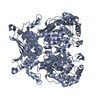
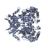
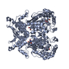
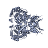
| Entry | Database: PDB / ID: 3if5 | |||||||||
|---|---|---|---|---|---|---|---|---|---|---|
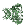
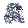
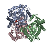
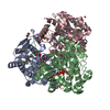
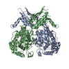
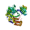
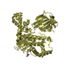
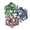
| Title | Crystal Structure Analysis of Mglu | |||||||||
 Components Components | Salt-tolerant glutaminase | |||||||||
 Keywords Keywords |  HYDROLASE / Fragment HYDROLASE / Fragment | |||||||||
| Function / homology |  Function and homology information Function and homology information | |||||||||
| Biological species |  Micrococcus luteus (bacteria) Micrococcus luteus (bacteria) | |||||||||
| Method | X-RAY DIFFRACTION / SYNCHROTRON / MAD / Resolution: 2.44 Å | |||||||||
 Authors Authors | Yoshimune, K. / Shirakihara, Y. | |||||||||
 Citation Citation | Journal: Febs J. / Year: 2010 Title: Crystal structure of salt-tolerant glutaminase from Micrococcus luteus K-3 in the presence and absence of its product L-glutamate and its activator Tris. Authors: Yoshimune, K. / Shirakihara, Y. / Wakayama, M. / Yumoto, I. #1: Journal: Biochem.Biophys.Res.Commun. / Year: 2006 Title: Crystal structure of a major fragment of the salt-tolerant glutaminase from Micrococcus luteus K-3 Authors: Yoshimune, K. / Shirakihara, Y. / Shiratori, A. / Wakayama, M. / Chantawannakul, P. / Moriguchi, M. | |||||||||
| History |
|
- Structure visualization
| Structure viewer | Molecule: MolmilJmol/JSmol |
|---|
- Downloads & links
-Download
| PDBx/mmCIF format | 3if5.cif.gz | 89.5 KB | Display | PDBx/mmCIF format |
|---|---|---|---|---|
| PDB format | pdb3if5.ent.gz | 68 KB | Display | PDB format |
| PDBx/mmJSON format | 3if5.json.gz | Tree view | PDBx/mmJSON format | |
| Others |  Other downloads Other downloads |
-Validation report
| Arichive directory | https://data.pdbj.org/pub/pdb/validation_reports/if/3if5ftp://data.pdbj.org/pub/pdb/validation_reports/if/3if5 | HTTPS FTP |
|---|
-Related structure data


-Links
 PDBj
PDBj
- Assembly
| Deposited unit | 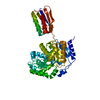
| ||||||||
|---|---|---|---|---|---|---|---|---|---|
| 1 | 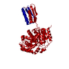
| ||||||||
| Unit cell |
|
-Components
| #1: Protein | Mass: 48303.516 Da / Num. of mol.: 1 Source method: isolated from a genetically manipulated source Source: (gene. exp.) Micrococcus luteus (bacteria) / Strain: K-3 / Gene: Glutaminase / Plasmid: pKK223-3 / Production host: Escherichia coli (E. coli) / Strain (production host): JM109 / References: UniProt: Q4U1A6, glutaminase |
|---|---|
| #2: Water | ChemComp-HOH / Water Mass: 18.015 Da / Num. of mol.: 115 / Source method: isolated from a natural source / Formula: H2O Mass: 18.015 Da / Num. of mol.: 115 / Source method: isolated from a natural source / Formula: H2O |
-Experimental details
-Experiment
| Experiment | Method: X-RAY DIFFRACTION / Number of used crystals: 1 |
|---|
- Sample preparation
| Crystal | Density Matthews: 2.52 Å3/Da / Density % sol: 51.18 % |
|---|---|
| Crystal grow | Temperature: 298 K / Method: vapor diffusion, hanging drop / pH: 7.5 Details: 700mM Sodium Citrate, 50mM HEPES, 5mM MgCl2, 5% Glycerol, pH 7.5, vapor diffusion, hanging drop, temperature 298K |
-Data collection
| Diffraction | Mean temperature: 100 K | ||||||||||||
|---|---|---|---|---|---|---|---|---|---|---|---|---|---|
| Diffraction source | Source: SYNCHROTRON / Site: SPring-8  / Beamline: BL41XU / Wavelength: 0.9791, 0.9793, 0.9700 / Beamline: BL41XU / Wavelength: 0.9791, 0.9793, 0.9700 | ||||||||||||
| Detector | Type: ADSC QUANTUM 4 / Detector: CCD / Date: Sep 26, 2001 | ||||||||||||
| Radiation | Monochromator: Rotated-inclined double-crystal monochromator Protocol: MAD / Monochromatic (M) / Laue (L): M / Scattering type: x-ray | ||||||||||||
| Radiation wavelength |
| ||||||||||||
| Reflection | Resolution: 2.4→36.18 Å / Num. all: 18367 / Num. obs: 17155 / Redundancy: 6.8 % / Biso Wilson estimate: 38.9 Å2 |
- Processing
| Software |
| ||||||||||||||||||||||||||||||||||||
|---|---|---|---|---|---|---|---|---|---|---|---|---|---|---|---|---|---|---|---|---|---|---|---|---|---|---|---|---|---|---|---|---|---|---|---|---|---|
| Refinement | Method to determine structure: MAD / Resolution: 2.44→36.18 Å / Rfactor Rfree error: 0.009 / Occupancy max: 1 / Occupancy min: 0.67 / Data cutoff high absF: 6969453 / Data cutoff low absF: 0 / Isotropic thermal model: RESTRAINED / Cross valid method: THROUGHOUT / σ(F): 0 / Details: BULK SOLVENT MODEL USED
| ||||||||||||||||||||||||||||||||||||
| Solvent computation | Solvent model: FLAT MODEL / Bsol: 46.196 Å2 / ksol: 0.4 e/Å3 | ||||||||||||||||||||||||||||||||||||
| Displacement parameters | Biso max: 110.85 Å2 / Biso mean: 39.072 Å2 / Biso min: 2.42 Å2
| ||||||||||||||||||||||||||||||||||||
| Refine analyze |
| ||||||||||||||||||||||||||||||||||||
| Refinement step | Cycle: LAST / Resolution: 2.44→36.18 Å
| ||||||||||||||||||||||||||||||||||||
| Refine LS restraints |
| ||||||||||||||||||||||||||||||||||||
| LS refinement shell | Resolution: 2.4→2.55 Å / Rfactor Rfree error: 0.034 / Total num. of bins used: 6
| ||||||||||||||||||||||||||||||||||||
| Xplor file |
|
