 Movie
Movie Controller


+ Open data

- Basic information
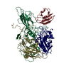
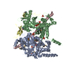
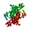
| Entry | Database: PDB / ID: 3icr | ||||||
|---|---|---|---|---|---|---|---|
| Title | Crystal structure of oxidized Bacillus anthracis CoADR-RHD | ||||||
 Components Components | Coenzyme A-Disulfide Reductase | ||||||
 Keywords Keywords |  OXIDOREDUCTASE / Pyridine nucleotide-disulfide oxidoreductase class I / Rhodanese / Coenzyme A / Flavin Adenine Dinucleotide OXIDOREDUCTASE / Pyridine nucleotide-disulfide oxidoreductase class I / Rhodanese / Coenzyme A / Flavin Adenine Dinucleotide | ||||||
| Function / homology |  Function and homology information Function and homology informationCoA-disulfide reductase (NADPH) activity / protein disulfide isomerase activity / NADP binding / flavin adenine dinucleotide bindingSimilarity search - Function | ||||||
| Biological species |  Bacillus anthracis (anthrax bacterium) Bacillus anthracis (anthrax bacterium) | ||||||
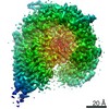
| Method | X-RAY DIFFRACTION / MOLECULAR REPLACEMENT / Resolution: 2.1 Å | ||||||
 Authors Authors | Wallen, J.R. / Claiborne, A. | ||||||
 Citation Citation | Journal: Biochemistry / Year: 2009 Title: Crystal structure and catalytic properties of Bacillus anthracis CoADR-RHD: implications for flavin-linked sulfur trafficking. Authors: Wallen, J.R. / Mallett, T.C. / Boles, W. / Parsonage, D. / Furdui, C.M. / Karplus, P.A. / Claiborne, A. | ||||||
| History |
|
- Structure visualization
| Structure viewer | Molecule: MolmilJmol/JSmol |
|---|
- Downloads & links
-Download
| PDBx/mmCIF format | 3icr.cif.gz | 246.6 KB | Display | PDBx/mmCIF format |
|---|---|---|---|---|
| PDB format | pdb3icr.ent.gz | 196.2 KB | Display | PDB format |
| PDBx/mmJSON format | 3icr.json.gz | Tree view | PDBx/mmJSON format | |
| Others |  Other downloads Other downloads |
-Validation report
| Arichive directory | https://data.pdbj.org/pub/pdb/validation_reports/ic/3icrftp://data.pdbj.org/pub/pdb/validation_reports/ic/3icr | HTTPS FTP |
|---|
-Related structure data


-Links
 PDBj
PDBj


- Assembly
| Deposited unit | 
| ||||||||
|---|---|---|---|---|---|---|---|---|---|
| 1 |
| ||||||||
| Unit cell |
|
-Components
| #1: Protein | Mass: 66008.312 Da / Num. of mol.: 2 Source method: isolated from a genetically manipulated source Source: (gene. exp.) Bacillus anthracis (anthrax bacterium) / Strain: Ames / Gene: BA0774, BAS0736, BA_0774, GBAA_0774 / Plasmid: pTHCm / Production host: Escherichia coli (E. coli) / Strain (production host): B834(DE3) / References: UniProt: Q81UT5, UniProt: A0A6L7H7X4*PLUS#2: Chemical | Flavin adenine dinucleotide  Mass: 785.550 Da / Num. of mol.: 2 / Source method: obtained synthetically / Formula: C27H33N9O15P2 / Comment: FAD*YM Mass: 785.550 Da / Num. of mol.: 2 / Source method: obtained synthetically / Formula: C27H33N9O15P2 / Comment: FAD*YM#3: Chemical | Coenzyme A  Mass: 767.534 Da / Num. of mol.: 2 / Source method: obtained synthetically / Formula: C21H36N7O16P3S Mass: 767.534 Da / Num. of mol.: 2 / Source method: obtained synthetically / Formula: C21H36N7O16P3S#4: Water | ChemComp-HOH / | Water Mass: 18.015 Da / Num. of mol.: 665 / Source method: isolated from a natural source / Formula: H2O Mass: 18.015 Da / Num. of mol.: 665 / Source method: isolated from a natural source / Formula: H2O |
|---|
-Experimental details
-Experiment
| Experiment | Method: X-RAY DIFFRACTION / Number of used crystals: 1 |
|---|
- Sample preparation
| Crystal | Density Matthews: 2.21 Å3/Da / Density % sol: 44.38 % |
|---|---|
| Crystal grow | Temperature: 288 K / Method: vapor diffusion, sitting drop / pH: 6 Details: 8-16% PEG 8000, 2% 2-methyl-2,4-pentanediol, 0.2M potassium acetate, pH 6.0, VAPOR DIFFUSION, SITTING DROP, temperature 288K |
-Data collection
| Diffraction | Mean temperature: 100 K |
|---|---|
| Diffraction source | Source: ROTATING ANODE / Type: RIGAKU MICROMAX-007 / Wavelength: 1.5418 Å |
| Detector | Type: RIGAKU SATURN 92 / Detector: CCD / Details: Confocal Blue Max-Flux |
| Radiation | Monochromator: graphite / Protocol: SINGLE WAVELENGTH / Monochromatic (M) / Laue (L): M / Scattering type: x-ray |
| Radiation wavelength | Wavelength: 1.5418 Å / Relative weight: 1 |
| Reflection | Resolution: 2.1→42.56 Å / Num. all: 66890 / Num. obs: 66422 / % possible obs: 99.3 % / Observed criterion σ(F): 0 / Observed criterion σ(I): 0 / Rmerge(I) obs: 0.097 / Net I/σ(I): 33.6 |
| Reflection shell | Resolution: 2.1→2.154 Å / Rmerge(I) obs: 0.302 / Mean I/σ(I) obs: 7.5 / Num. unique all: 61873 / % possible all: 92.5 |
- Processing
| Software |
| ||||||||||||||||||||||||||||||||||||||||||||||||||||||||||||||||||||||||||||||||||||||||||
|---|---|---|---|---|---|---|---|---|---|---|---|---|---|---|---|---|---|---|---|---|---|---|---|---|---|---|---|---|---|---|---|---|---|---|---|---|---|---|---|---|---|---|---|---|---|---|---|---|---|---|---|---|---|---|---|---|---|---|---|---|---|---|---|---|---|---|---|---|---|---|---|---|---|---|---|---|---|---|---|---|---|---|---|---|---|---|---|---|---|---|---|
| Refinement | Method to determine structure: MOLECULAR REPLACEMENT Starting model: 3 wavelength MAD structure deteremined on Selenomethionine-containing CoADR-RHD Resolution: 2.1→42.56 Å / Cor.coef. Fo:Fc: 0.953 / Cor.coef. Fo:Fc free: 0.926 / SU B: 8.74 / SU ML: 0.126 / TLS residual ADP flag: LIKELY RESIDUAL / Cross valid method: THROUGHOUT / ESU R: 0.229 / ESU R Free: 0.182 / Stereochemistry target values: MAXIMUM LIKELIHOOD
| ||||||||||||||||||||||||||||||||||||||||||||||||||||||||||||||||||||||||||||||||||||||||||
| Solvent computation | Ion probe radii: 0.8 Å / Shrinkage radii: 0.8 Å / VDW probe radii: 1.4 Å / Solvent model: BABINET MODEL WITH MASK | ||||||||||||||||||||||||||||||||||||||||||||||||||||||||||||||||||||||||||||||||||||||||||
| Displacement parameters | Biso mean: 23.417 Å2
| ||||||||||||||||||||||||||||||||||||||||||||||||||||||||||||||||||||||||||||||||||||||||||
| Refinement step | Cycle: LAST / Resolution: 2.1→42.56 Å
| ||||||||||||||||||||||||||||||||||||||||||||||||||||||||||||||||||||||||||||||||||||||||||
| Refine LS restraints |
| ||||||||||||||||||||||||||||||||||||||||||||||||||||||||||||||||||||||||||||||||||||||||||
| LS refinement shell | Resolution: 2.1→2.154 Å / Total num. of bins used: 20
| ||||||||||||||||||||||||||||||||||||||||||||||||||||||||||||||||||||||||||||||||||||||||||
| Refinement TLS params. | Method: refined / Refine-ID: X-RAY DIFFRACTION
| ||||||||||||||||||||||||||||||||||||||||||||||||||||||||||||||||||||||||||||||||||||||||||
| Refinement TLS group |
|
